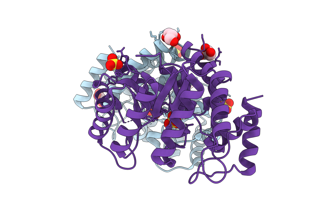
1F6K image
Entry Detail
PDB ID:
1F6K
Keywords:
Title:
CRYSTAL STRUCTURE ANALYSIS OF N-ACETYLNEURAMINATE LYASE FROM HAEMOPHILUS INFLUENZAE: CRYSTAL FORM II
Biological Source:
Source Organism:
Host Organism:
PDB Version:
Deposition Date:
2000-06-21
Release Date:
2000-12-20
Method Details:
Experimental Method:
Resolution:
1.60 Å
R-Value Free:
0.22
R-Value Work:
0.18
Space Group:
P 21 21 2
Macromolecular Entities
Polymer Type:polypeptide(L)
Description:N-ACETYLNEURAMINATE LYASE
Chain IDs:A, B (auth: C)
Chain Length:293
Number of Molecules:2
Biological Source:Haemophilus influenzae
Primary Citation
Active site modulation in the N-acetylneuraminate lyase sub-family as revealed by the structure of the inhibitor-complexed Haemophilus influenzae enzyme.
J.Mol.Biol. 303 405 421 (2000)
PMID: 11031117 DOI: 10.1006/jmbi.2000.4138

Abstact

The N-acetylneuraminate lyase (NAL) sub-family of (beta/alpha)(8) enzymes share a common catalytic step but catalyse reactions in different biological pathways. Known examples include NAL, dihydrodipicolinate synthetase (DHDPS), d-5-keto-4-deoxyglucarate dehydratase, 2-keto-3-deoxygluconate aldolase, trans-o-hydroxybenzylidenepyruvate hydrolase-aldolase and trans-2'-carboxybenzalpyruvate hydratase-aldolase. Little is known about the way in which the three-dimensional structure of the respective active sites are modulated across the sub-family to achieve cognate substrate recognition. We present here the structure of Haemophilus influenzae NAL determined by X-ray crystallography to a maximum resolution of 1.60 A, in native form and in complex with three substrate analogues (sialic acid alditol, 4-deoxy-sialic acid and 4-oxo-sialic acid). These structures reveal for the first time the mode of binding of the complete substrate in the NAL active site. On the basis of the above structures, that of substrate-complexed DHDPS and sequence comparison across the sub-family we are able to propose a unified model for active site modulation. The model is one of economy, allowing wherever appropriate the retention or relocation of residues associated with binding common substrate substituent groups. Our structures also suggest a role for the strictly conserved tyrosine residue found in all active sites of the sub-family, namely that it mediates proton abstraction by the alpha-keto acid carboxylate in a substrate-assisted catalytic reaction pathway.

Legend

Protein

Chemical

Disease

Primary Citation of related structures