1F5S image
Deposition Date 2000-06-15
Release Date 2001-06-20
Last Version Date 2024-03-13
Entry Detail
PDB ID:
1F5S
Keywords:
Title:
CRYSTAL STRUCTURE OF PHOSPHOSERINE PHOSPHATASE FROM METHANOCOCCUS JANNASCHII
Biological Source:
Source Organism:
Host Organism:
Method Details:
Experimental Method:
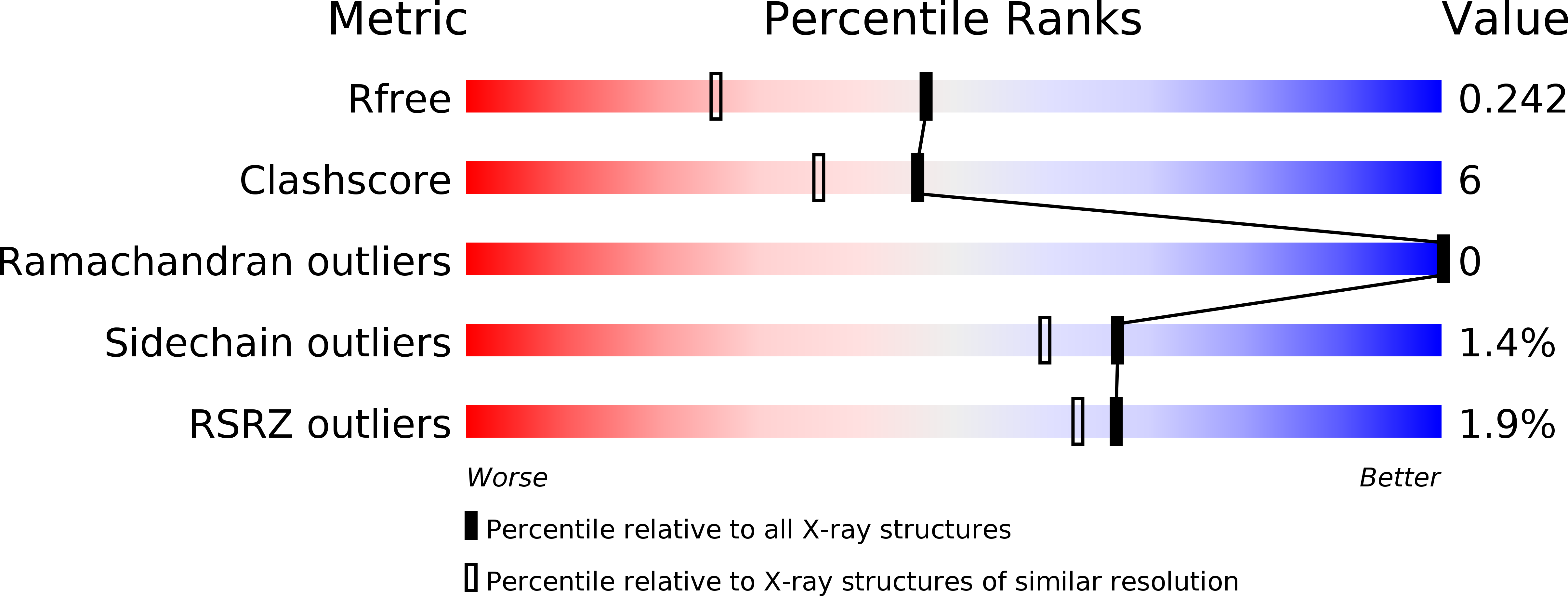
Resolution:
1.80 Å
R-Value Free:
0.23
R-Value Work:
0.19
R-Value Observed:
0.20
Space Group:
P 21 21 2
Macromolecular Entities
Structures with similar UniProt ID
Protein Blast
Polymer Type:polypeptide(L)
Molecule:PHOSPHOSERINE PHOSPHATASE (PSP)
Gene (Uniprot):MJ1594
Chain IDs:A, B
Chain Length:211
Number of Molecules:2
Biological Source:Methanocaldococcus jannaschii
Primary Citation
Crystal structure of phosphoserine phosphatase from Methanococcus jannaschii, a hyperthermophile, at 1.8 A resolution.
Structure 9 65 72 (2001)
PMID: 11342136 DOI: 10.1016/S0969-2126(00)00558-X

Abstact

BACKGROUND: D-Serine is a co-agonist of the N-methyl-D-aspartate subtype of glutamate receptors, a major neurotransmitter receptor family in mammalian nervous systems. D-Serine is converted from L-serine, 90% of which is the product of the enzyme phosphoserine phosphatase (PSP). PSP from M. jannaschii (MJ) shares significant sequence homology with human PSP. PSPs and P-type ATPases are members of the haloacid dehalogenase (HAD)-like hydrolase family, and all members share three conserved sequence motifs. PSP and P-type ATPases utilize a common mechanism that involves Mg(2+)-dependent phosphorylation and autodephosphorylation at an aspartyl side chain in the active site. The strong resemblance in sequence and mechanism implies structural similarity among these enzymes. RESULTS: The PSP crystal structure resembles the NAD(P) binding Rossmann fold with a large insertion of a four-helix-bundle domain and a beta hairpin. Three known conserved sequence motifs are arranged next to each other in space and outline the active site. A phosphate and a magnesium ion are bound to the active site. The active site is within a closed environment between the core alpha/beta domain and the four-helix-bundle domain. CONCLUSIONS: The crystal structure of MJ PSP was determined at 1.8 A resolution. Critical residues were assigned based on the active site structure and ligand binding geometry. The PSP structure is in a closed conformation that may resemble the phosphoserine bound state or the state after autodephosphorylation. Compared to a P-type ATPase (Ca(2+)-ATPase) structure, which is in an open state, this PSP structure appears also to be a good model for the closed conformation of P-type ATPase.

Legend

Protein

Chemical

Disease

Primary Citation of related structures