Deposition Date
2000-06-13
Release Date
2000-06-28
Last Version Date
2024-02-07
Entry Detail
PDB ID:
1F5C
Keywords:
Title:
CRYSTAL STRUCTURE OF F25H FERREDOXIN 1 MUTANT FROM AZOTOBACTER VINELANDII AT 1.75 ANGSTROM RESOLUTION
Biological Source:
Source Organism(s):
Azotobacter vinelandii (Taxon ID: 354)
Expression System(s):
Method Details:
Experimental Method:
Resolution:
1.75 Å
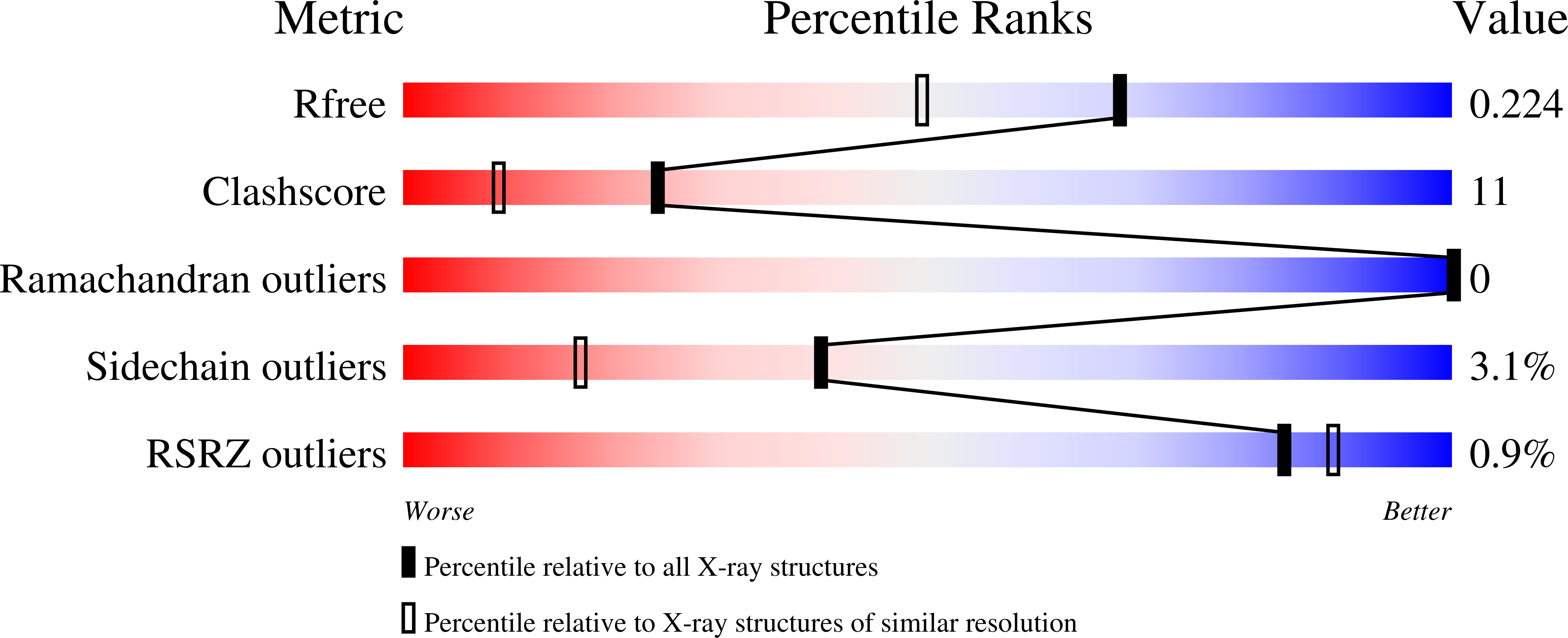
R-Value Free:
0.22
R-Value Work:
0.20
R-Value Observed:
0.20
Space Group:
P 41 21 2