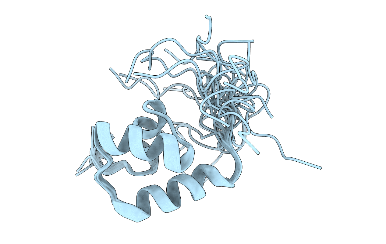
1F43 image
Entry Detail
PDB ID:
1F43
Keywords:
Title:
SOLUTION STRUCTURE OF THE MATA1 HOMEODOMAIN
Biological Source:
Source Organism:
Host Organism:
PDB Version:
Deposition Date:
2000-06-07
Release Date:
2000-07-26
Method Details:
Experimental Method:
Conformers Calculated:
100
Conformers Submitted:
20
Selection Criteria:
target function
Macromolecular Entities
Polymer Type:polypeptide(L)
Description:MATING-TYPE PROTEIN A-1
Chain IDs:A
Chain Length:61
Number of Molecules:1
Biological Source:Saccharomyces cerevisiae
Ligand Molecules
Primary Citation
Cooperative ordering in homeodomain-DNA recognition: solution structure and dynamics of the MATa1 homeodomain.
Biochemistry 39 10045 10054 (2000)
PMID: 10955992 DOI: 10.1021/bi000677z

Abstact

The mating type homeodomain proteins, MATa1 and MATalpha2, combine to form a heterodimer to bind DNA in diploid yeast cells. The a1-alpha2 heterodimer tightly and specifically binds haploid-specific gene operators to repress transcription. On its own, however, the a1 homeodomain does not bind DNA in a sequence-specific manner. To help understand this interaction, we describe the solution structure and backbone dynamics of the free a1 homeodomain. Free a1 in solution is an ensemble of structures having flexible hinges at the two turns in the small protein fold. Conformational changes in the a1 homeodomain upon ternary complex formation are located in the loop between helix 1 and helix 2, where the C-terminal tail of alpha2 binds to form the heterodimer, and at the C-terminus of helix 3, the DNA recognition helix. The observed differences, comparing the free and bound a1 structures, suggest a mechanism linking van der Waals stacking changes to the ordering of a final turn in the DNA-binding helix of a1. The tail of alpha2 induces changes in loop 1 of a1 that push it toward a properly folded DNA binding conformation.

Legend

Protein

Chemical

Disease

Primary Citation of related structures