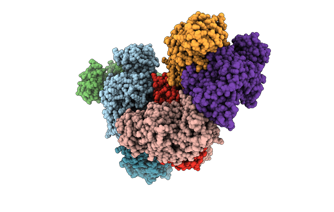
1F3W image
Entry Detail
PDB ID:
1F3W
Keywords:
Title:
RECOMBINANT RABBIT MUSCLE PYRUVATE KINASE
Biological Source:
Source Organism:
Host Organism:
PDB Version:
Deposition Date:
2000-06-06
Release Date:
2001-10-03
Method Details:
Experimental Method:
Resolution:
3.00 Å
R-Value Free:
0.25
R-Value Work:
0.23
R-Value Observed:
0.23
Space Group:
P 1
Macromolecular Entities
Polymer Type:polypeptide(L)
Description:PYRUVATE KINASE
Chain IDs:A, B, C, D, E, F, G, H
Chain Length:530
Number of Molecules:8
Biological Source:Oryctolagus cuniculus
Primary Citation
Structural and functional linkages between subunit interfaces in mammalian pyruvate kinase.
J.Mol.Biol. 312 525 540 (2001)
PMID: 11563914 DOI: 10.1006/jmbi.2001.4978

Abstact

Mammalian pyruvate kinase (PK) is a four-domain enzyme that is active as a homo-tetramer. Tissue-specific isozymes of PK exhibit distinct levels of allosteric regulation. PK expressed in muscle tissue (M1-PK) shows hyperbolic steady-state kinetics, whereas PK expressed in kidney tissue (M2-PK) displays sigmoidal kinetics. Rabbit M1 and M2-PK are isozymes whose sequences differ in only 22 out of 530 residues per subunit, and these changes are localized in an inter-subunit interface. Previous studies have shown that a single amino acid mutation to M1-PK at either the Y (S402P) or Z (T340 M) subunit interface can confer a level of allosteric regulation that is intermediate to M1-PK and M2-PK. In an effort to elucidate the roles of the inter-subunit interaction in signal transmission and the functional/structural connectivity between these interfaces, the S402P mutant of M1-PK was crystallized and its structure resolved to 2.8 A. Although the overall S402P M1-PK structure is nearly identical with the wild-type structure within experimental error, significant differences in the conformation of the backbone are found at the site of mutation along the Y interface. In addition, there is a significant change along the Z interface, namely, a loss of an inter-subunit salt-bridge between Asp177 of domain B and Arg341 of domain A of the opposing subunit. Concurrent with the loss of the salt-bridge is an increase in the degree of rotational flexibility of domain B that constitutes the active site. Comparison of previous PK structures shows a correlation between an increase in this domain movement with the loss of the Asp177: Arg341 salt-bridge. These results identify the structural linkages between the Y and Z interfaces in regulating the interconversion of conformational states of rabbit M1-PK.

Legend

Protein

Chemical

Disease

Primary Citation of related structures