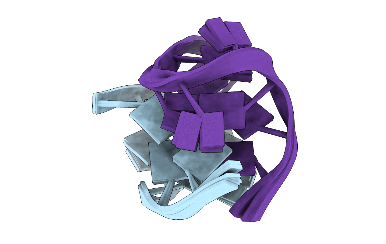
1F3S image
Deposition Date 2000-06-06
Release Date 2000-11-13
Last Version Date 2024-05-22
Entry Detail
PDB ID:
1F3S
Keywords:
Title:
Solution Structure of DNA Sequence GGGTTCAGG Forms GGGG Tetrade and G(C-A) Triad.
Biological Source:
Source Organism:
(Taxon ID: ) (Taxon ID: )
Method Details:
Experimental Method:
Conformers Calculated:
18
Conformers Submitted:
10
Selection Criteria:
structures with the lowest energy
Macromolecular Entities
Polymer Type:polydeoxyribonucleotide
Molecule:DNA (5'-D(*GP*GP*GP*TP*TP*CP*AP*GP*G)-3')
Chain IDs:A, B
Chain Length:9
Number of Molecules:2
Biological Source:
Ligand Molecules
Primary Citation
A two-stranded template-based approach to G.(C-A) triad formation: designing novel structural elements into an existing DNA framework.
J.Mol.Biol. 301 129 146 (2000)
PMID: 10926497 DOI: 10.1006/jmbi.2000.3932

Abstact

We have designed a DNA sequence, d(G-G-G-T-T-C-A-G-G), which dimerizes to form a 2-fold symmetric G-quadruplex in which G(syn). G(anti).G(syn).G(anti) tetrads are sandwiched between all trans G. (C-A) triads. The NMR-based solution structural analysis was greatly aided by monitoring hydrogen bond alignments across N-H...N and N-H...O==C hydrogen bonds within the triad and tetrad, in a uniformly ((13)C,(15)N)-labeled sample of the d(G-G-G-T-T-C-A-G-G) sequence. The solution structure establishes that the guanine base-pairs with the cytosine through Watson-Crick G.C pair formation and with adenine through sheared G.A mismatch formation within the G.(C-A) triad. A model of triad DNA was constructed that contains the experimentally determined G.(C-A) triad alignment as the repeating stacked unit.

Legend

Protein

Chemical

Disease

Primary Citation of related structures