
Deposition Date
2000-06-01
Release Date
2000-07-26
Last Version Date
2024-10-30
Entry Detail
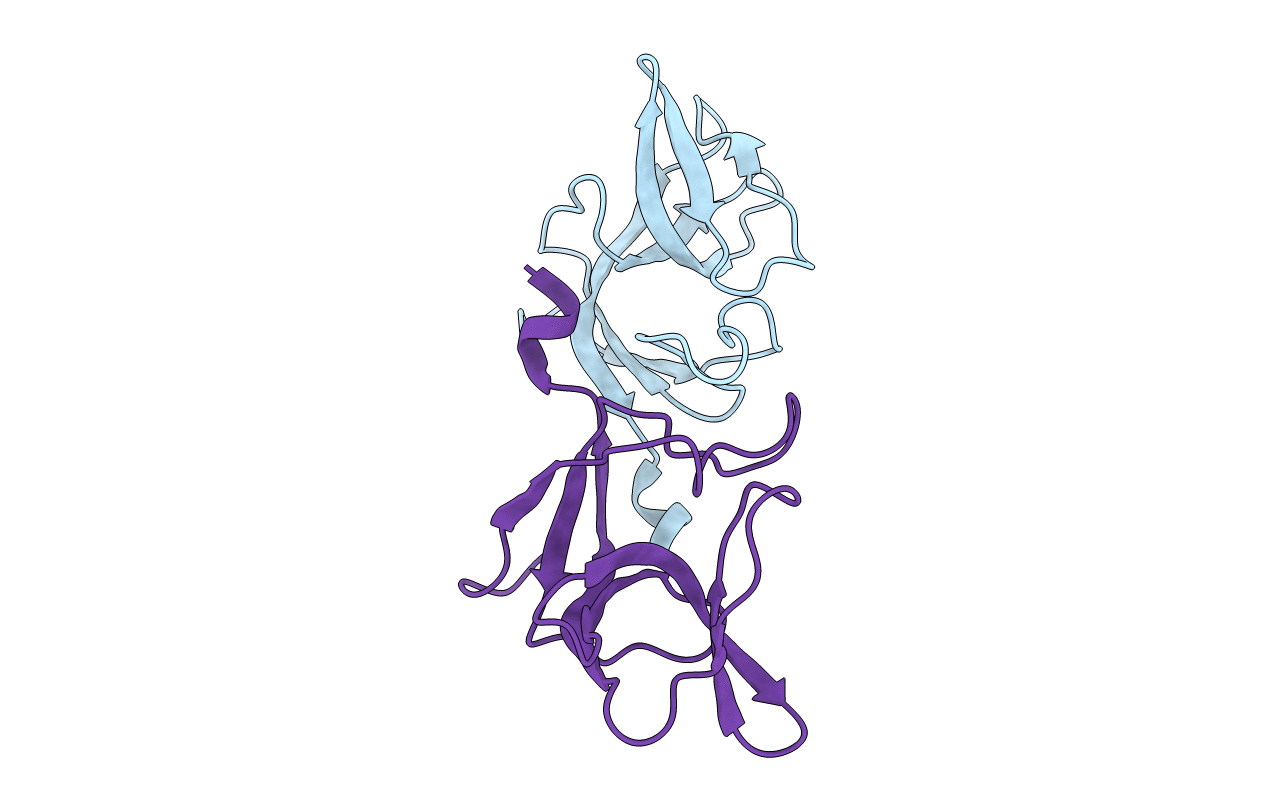
PDB ID:
1F39
Keywords:
Title:
CRYSTAL STRUCTURE OF THE LAMBDA REPRESSOR C-TERMINAL DOMAIN
Biological Source:
Source Organism(s):
Enterobacteria phage lambda (Taxon ID: 10710)
Expression System(s):
Method Details:
Experimental Method:
Resolution:
1.90 Å
R-Value Free:
0.25
R-Value Work:
0.22
R-Value Observed:
0.22
Space Group:
C 2 2 21
