1F2V image
Deposition Date 2000-05-29
Release Date 2001-07-18
Last Version Date 2024-02-07
Entry Detail
PDB ID:
1F2V
Keywords:
Title:
CRYSTAL STRUCTURE ANALYSIS OF PRECORRIN-8X METHYLMUTASE OF AEROBIC VITAMIN B12 SYNTHESIS
Biological Source:
Source Organism:
Host Organism:
Method Details:
Experimental Method:
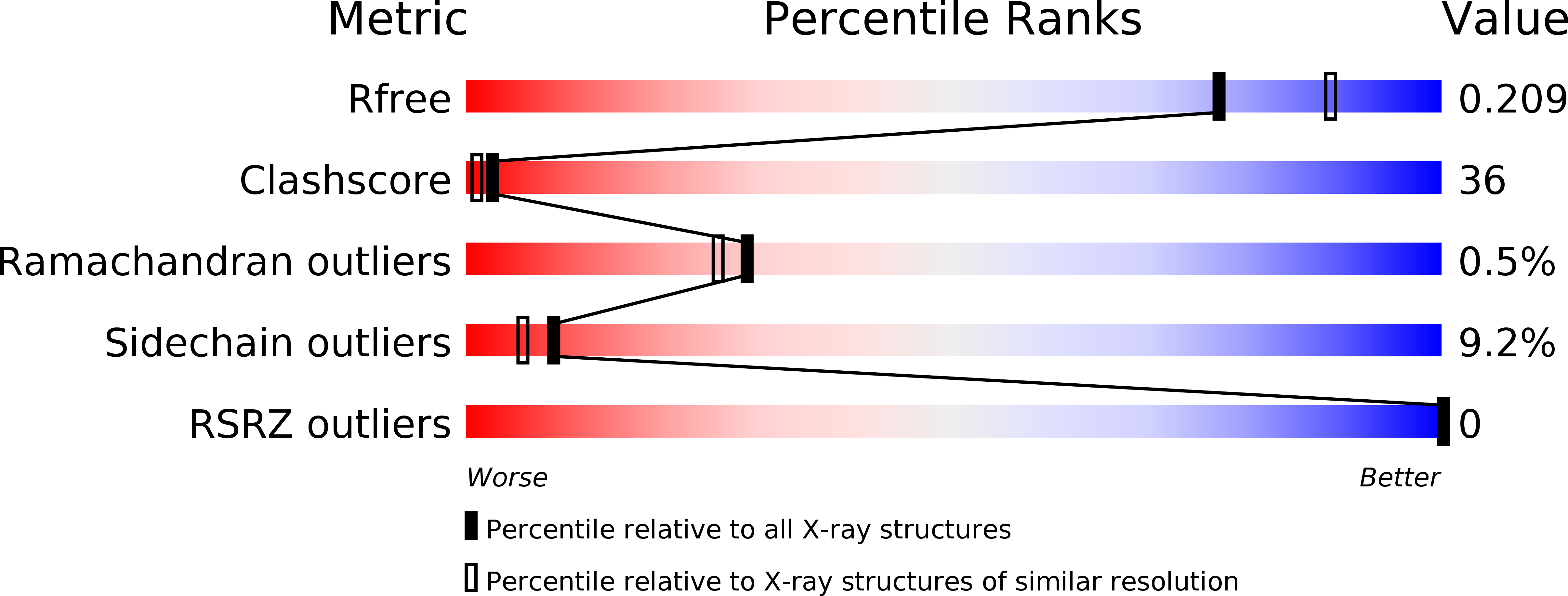
Resolution:
2.10 Å
R-Value Free:
0.23
R-Value Work:
0.18
R-Value Observed:
0.18
Space Group:
C 1 2 1
Macromolecular Entities
Polymer Type:polypeptide(L)
Molecule:PRECORRIN-8X METHYLMUTASE
Gene (Uniprot):cobH
Chain IDs:A
Chain Length:219
Number of Molecules:1
Biological Source:Pseudomonas denitrificans
Primary Citation
Crystal structure of precorrin-8x methyl mutase.
Structure 9 587 596 (2001)
PMID: 11470433 DOI: 10.1016/S0969-2126(01)00618-9

Abstact

BACKGROUND: The crystal structure of precorrin-8x methyl mutase (CobH), an enzyme of the aerobic pathway to vitamin B12, provides evidence that the mechanism for methyl migration can plausibly be regarded as an allowed [1,5]-sigmatropic shift of a methyl group from C-11 to C-12 at the C ring of precorrin-8x to afford hydrogenobyrinic acid. RESULTS: The dimeric structure of CobH creates a set of shared active sites that readily discriminate between different tautomers of precorrin-8x and select a discrete tautomer for sigmatropic rearrangement. The active site contains a strictly conserved histidine residue close to the site of methyl migration in ring C of the substrate. CONCLUSION: Analysis of the structure with bound product suggests that the [1,5]-sigmatropic shift proceeds by protonation of the ring C nitrogen, leading to subsequent methyl migration.

Legend

Protein

Chemical

Disease

Primary Citation of related structures