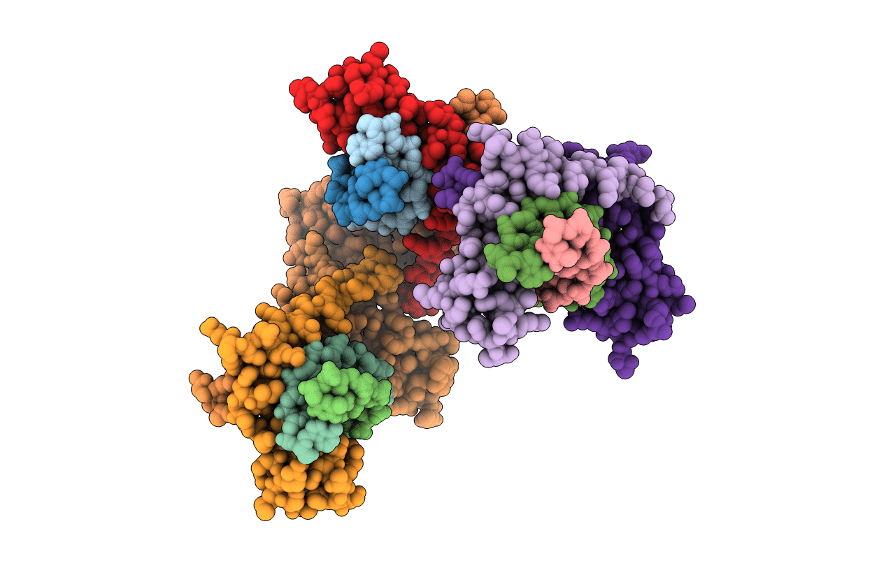
1F2I image
Deposition Date 2000-05-25
Release Date 2001-09-14
Last Version Date 2024-02-07
Entry Detail
PDB ID:
1F2I
Title:
COCRYSTAL STRUCTURE OF SELECTED ZINC FINGER DIMER BOUND TO DNA
Biological Source:
Source Organism:
Mus musculus (Taxon ID: 10090)
Host Organism:
Method Details:
Experimental Method:
Resolution:
2.35 Å
R-Value Free:
0.25
R-Value Work:
0.21
R-Value Observed:
0.21
Space Group:
P 31
Macromolecular Entities
Structures with similar UniProt ID
Protein Blast
Polymer Type:polypeptide(L)
Molecule:FUSION OF N-TERMINAL 17-MER PEPTIDE EXTENSION TO ZIF12
Gene (Uniprot):Egr1
Chain IDs:G, H, I, J, K, L
Chain Length:73
Number of Molecules:6
Biological Source:Mus musculus
Ligand Molecules
Primary Citation
Selected peptide extension contacts hydrophobic patch on neighboring zinc finger and mediates dimerization on DNA.
Nat.Struct.Biol. 8 589 593 (2001)
PMID: 11427887 DOI: 10.1038/89617

Abstact

Protein-protein interactions often play a crucial role in stabilizing protein-DNA complexes and thus facilitate site-specific DNA recognition. We have worked to incorporate such protein-protein contacts into our design and selection strategies for short peptide extensions that promote cooperative binding of zinc finger proteins to DNA. We have determined the crystal structure of one of these fusion protein-DNA complexes. The selected peptide extension was found to mediate dimerization by reaching across the dyad axis and contacting a hydrophobic patch on the surface of the zinc finger bound to the adjacent DNA site. The peptide-zinc finger protein interactions observed in this structure are similar to those of some homeodomain heterodimers. We also find that the region of the zinc finger surface contacted by the selected peptide extension corresponds to surfaces that also make key interactions in the zinc finger proteins GLI and SWI5.

Legend

Protein

Chemical

Disease

Primary Citation of related structures