1F0V image
Deposition Date 2000-05-17
Release Date 2001-02-21
Last Version Date 2024-11-20
Entry Detail
PDB ID:
1F0V
Keywords:
Title:
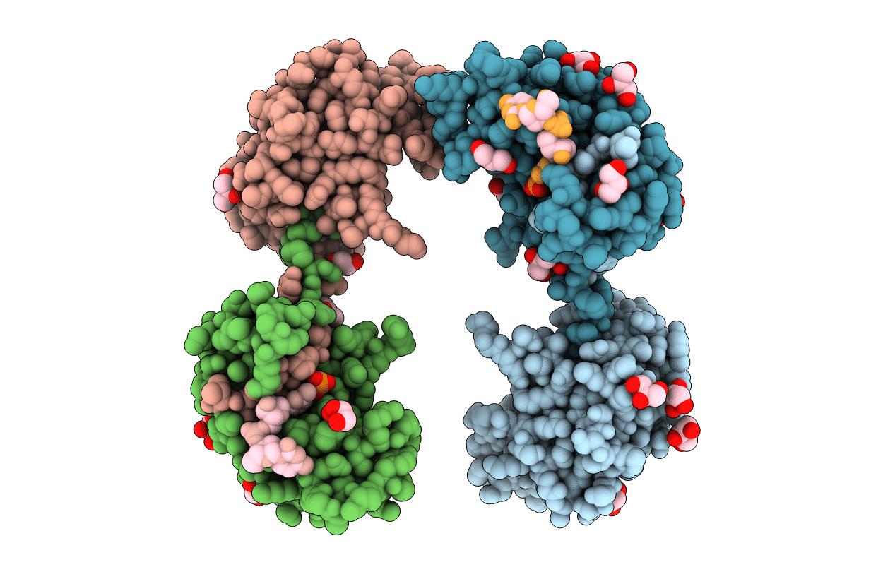
Crystal structure of an Rnase A dimer displaying a new type of 3D domain swapping
Biological Source:
Source Organism:
Bos taurus (Taxon ID: 9913)
Method Details:
Experimental Method:
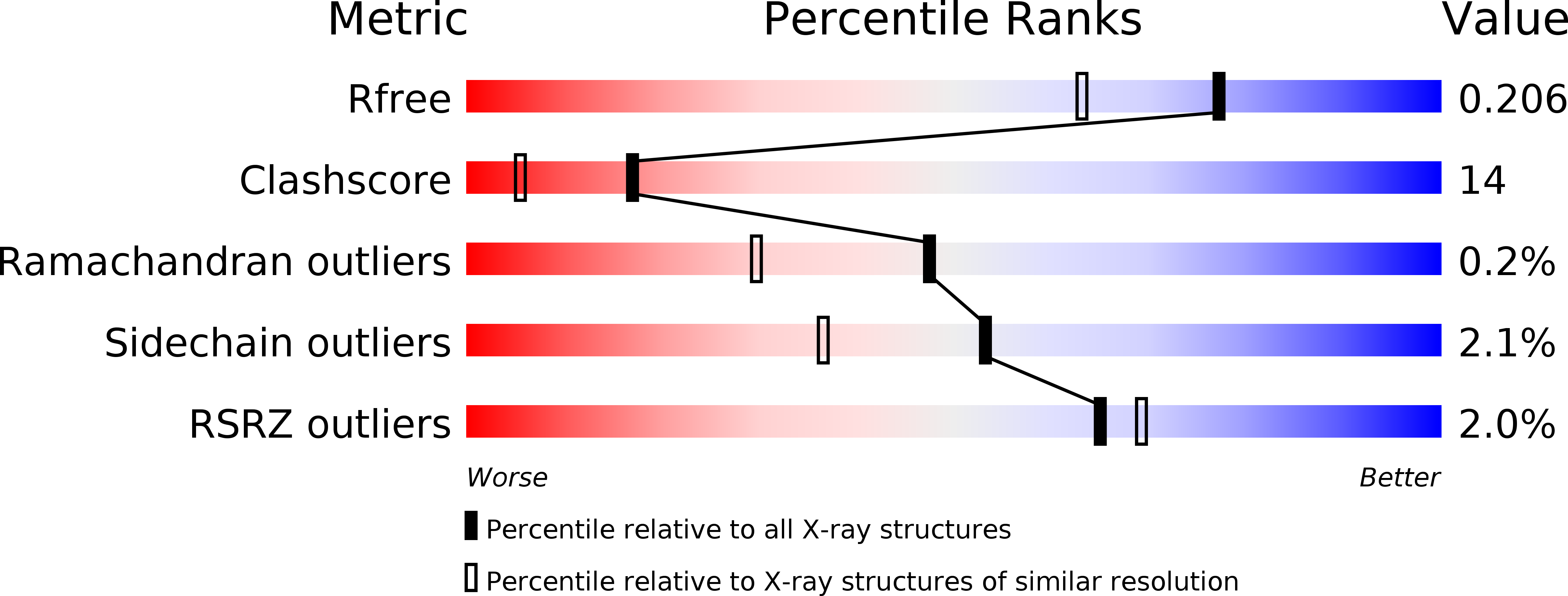
Resolution:
1.70 Å
R-Value Free:
0.21
R-Value Work:
0.18
Space Group:
P 1 21 1
Macromolecular Entities
Polymer Type:polypeptide(L)
Molecule:RIBONUCLEASE A
Gene (Uniprot):RNASE1
Chain IDs:E (auth: A), F (auth: B), G (auth: C), H (auth: D)
Chain Length:124
Number of Molecules:4
Biological Source:Bos taurus
Primary Citation
A domain-swapped RNase A dimer with implications for amyloid formation
Nat.Struct.Biol. 8 211 214 (2001)
PMID: 11224563 DOI: 10.1038/84941

Abstact

Bovine pancreatic ribonuclease (RNase A) forms two types of dimers (a major and a minor component) upon concentration in mild acid. These two dimers exhibit different biophysical and biochemical properties. Earlier we reported that the minor dimer forms by swapping its N-terminal alpha-helix with that of an identical molecule. Here we find that the major dimer forms by swapping its C-terminal beta-strand, thus revealing the first example of three-dimensional (3D) domain swapping taking place in different parts of the same protein. This feature permits RNase A to form tightly bonded higher oligomers. The hinge loop of the major dimer, connecting the swapped beta-strand to the protein core, resembles a short segment of the polar zipper proposed by Perutz and suggests a model for aggregate formation by 3D domain swapping with a polar zipper.

Legend

Protein

Chemical

Disease

Primary Citation of related structures