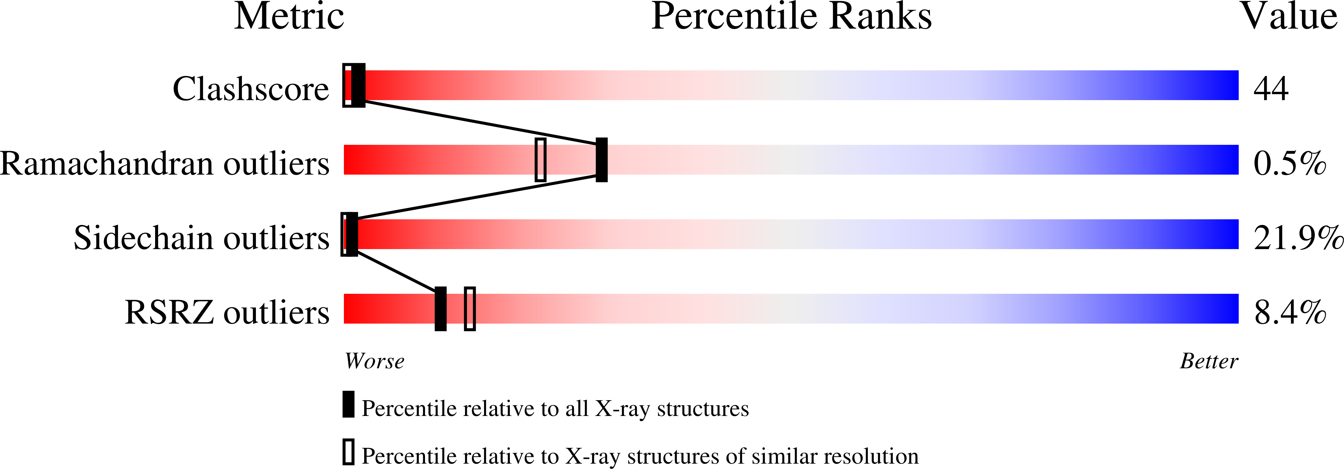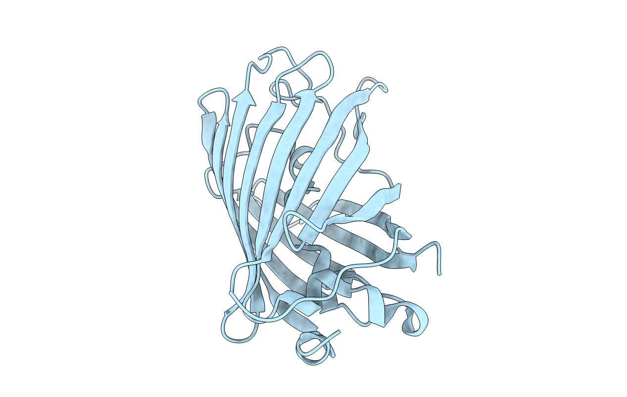
1F09 image
Deposition Date 2000-05-15
Release Date 2000-11-17
Last Version Date 2024-10-16
Entry Detail
PDB ID:
1F09
Title:
CRYSTAL STRUCTURE OF THE GREEN FLUORESCENT PROTEIN (GFP) VARIANT YFP-H148Q WITH TWO BOUND IODIDES
Biological Source:
Source Organism:
Host Organism:
Method Details:
Experimental Method:
Resolution:
2.14 Å
R-Value Work:
0.20
R-Value Observed:
0.20
Space Group:
P 21 21 21
Macromolecular Entities
Polymer Type:polypeptide(L)
Molecule:GREEN FLUORESCENT PROTEIN
Gene (Uniprot):GFP
Mutations:S65G, V68L, S72A, Q80R, T203Y, H148Q
Chain IDs:A
Chain Length:238
Number of Molecules:1
Biological Source:Aequorea victoria
Modified Residue
Compound ID Chain ID Parent Comp ID Details 2D Image
CR2 A GLY ?
Ligand Molecules
Primary Citation
Crystallographic and energetic analysis of binding of selected anions to the yellow variants of green fluorescent protein.
J.Mol.Biol. 301 157 171 (2000)
PMID: 10926499 DOI: 10.1006/jmbi.2000.3905

Abstact

The fluorescence emission of yellow fluorescent proteins (YFPs) has been shown to respond rapidly and reversibly to changes in the concentration of some small anions such as halides; this allows for the use of YFPs as genetically encodable Cl(-) sensors that may be targeted to specific organelles in living cells. Fluorescence is suppressed due to protonation of the chromophore upon anion binding, with a stronger level of interaction at low pH values. At pH 6.0, the apparent dissociation constant (K(app)) for Cl(-) is 32 mM for YFP and 22 mM for YFP-H148Q, whereas at pH 7.5, K(app) is 777 mM and 154 mM, respectively. In the cytosol, YFP-H148Q appears most promising as a halide sensor due to its high degree of sensitivity towards I(-) (K(app)=23 mM at pH 7.5). To aid in the design of variants with improved levels of specificity and affinity for Cl(-), we solved apo and I(-)-bound crystal structures of YFP-H148Q to 2.1 A resolution. The halide-binding site is found near van der Waals contact with the chromophore imidazolinone oxygen atom, in a small buried cavity adjacent to Arg96, which provides electrostatic stabilization. The halide ion is hydrogen bonded to the phenol group of T203Y, consistent with a mutational analysis that indicates that T203Y is indispensible for tight binding. A series of conformational changes occurs in the amphiphilic site upon anion binding, which appear to be propagated to the beta-bulge region around residue 148 on the protein surface. Anion binding raises the chromophore pK(a) values, since delocalization of the phenolate negative charge over the chromophore skeleton is suppressed. Extraction of microscopic binding constants for the linked equilibrium between anion and proton binding indicates that anion selectivity by YFP is related to hydration forces. Specific suggestions to improve Cl(-) binding to YFP-H148Q based on size and hydration energy are proposed.

Legend

Protein

Chemical

Disease

Primary Citation of related structures