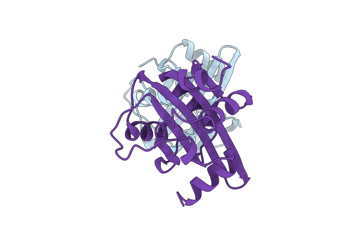
1F08 image
Deposition Date 2000-05-15
Release Date 2001-05-16
Last Version Date 2024-02-07
Entry Detail
PDB ID:
1F08
Keywords:
Title:
CRYSTAL STRUCTURE OF THE DNA-BINDING DOMAIN OF THE REPLICATION INITIATION PROTEIN E1 FROM PAPILLOMAVIRUS
Biological Source:
Source Organism:
Host Organism:
Method Details:
Experimental Method:
Resolution:
1.90 Å
R-Value Free:
0.27
R-Value Work:
0.24
Space Group:
P 21 21 21
Macromolecular Entities
Polymer Type:polypeptide(L)
Molecule:REPLICATION PROTEIN E1
Gene (Uniprot):E1
Chain IDs:A, B
Chain Length:148
Number of Molecules:2
Biological Source:Bovine papillomavirus
Ligand Molecules
Primary Citation
Crystal structure of the DNA binding domain of the replication initiation protein E1 from papillomavirus.
Mol.Cell 6 149 158 (2000)
PMID: 10949036 DOI: 10.1016/S1097-2765(00)00016-2

Abstact

Papillomaviral infection causes both benign and malignant lesions and is a necessary cause of cervical carcinoma. Replication of this virus requires the replication initiation proteins E1 and E2, which bind cooperatively at the origin of replication (ori) as an (E1)2-(E2)2-DNA complex. This is a precursor to larger E1 complexes that distort and unwind the ori. We present the crystal structure of the E1 DNA binding domain refined to 1.9 A resolution. Residues critical for DNA binding are located on an extended loop and an alpha helix. We identify the E1 dimerization surface by selective mutations at an E1/E1 interface observed in the crystal and propose a model for the (E1)2-DNA complex. These and other observations suggest how the E1 DNA binding domain orchestrates assembly of the hexameric helicase on the ori.

Legend

Protein

Chemical

Disease

Primary Citation of related structures