
Deposition Date
2000-05-11
Release Date
2000-06-23
Last Version Date
2024-11-13
Entry Detail
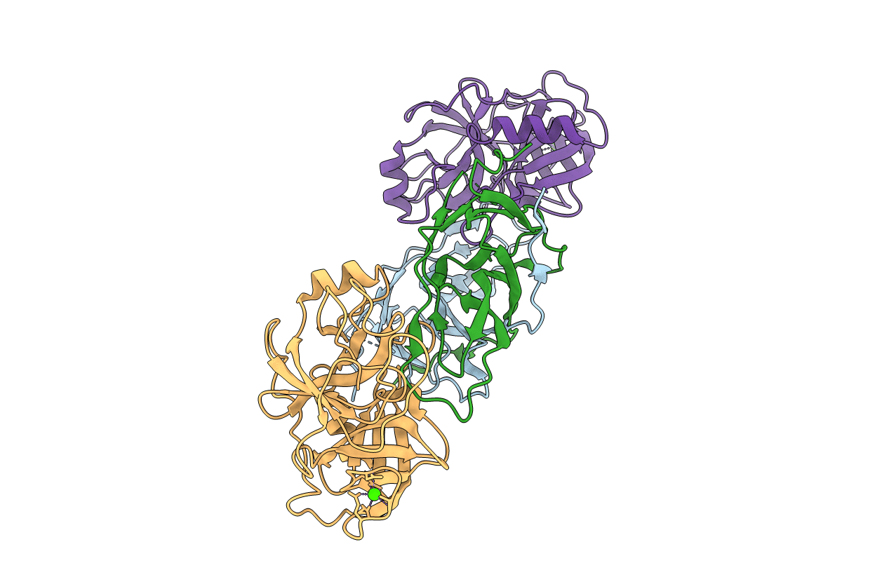
PDB ID:
1EZS
Keywords:
Title:
CRYSTAL STRUCTURE OF ECOTIN MUTANT M84R, W67A, G68A, Y69A, D70A BOUND TO RAT ANIONIC TRYPSIN II
Biological Source:
Source Organism:
Escherichia coli (Taxon ID: 562)
Rattus norvegicus (Taxon ID: 10116)
Rattus norvegicus (Taxon ID: 10116)
Host Organism:
Method Details:
Experimental Method:
Resolution:
2.30 Å
R-Value Free:
0.23
R-Value Work:
0.18
R-Value Observed:
0.18
Space Group:
P 1
