1EYB image
Deposition Date 2000-05-05
Release Date 2000-11-05
Last Version Date 2024-11-13
Entry Detail
PDB ID:
1EYB
Keywords:
Title:
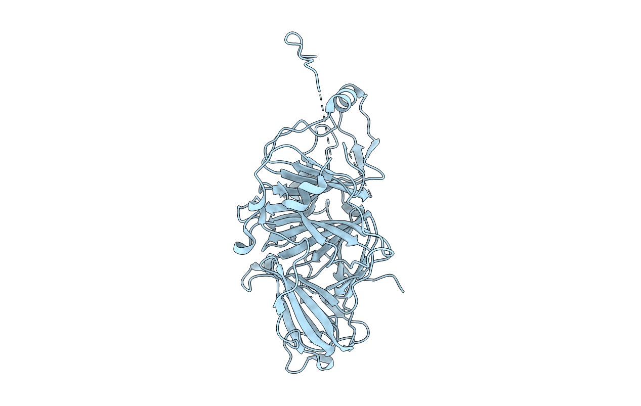
CRYSTAL STRUCTURE OF APO HUMAN HOMOGENTISATE DIOXYGENASE
Biological Source:
Source Organism:
Homo sapiens (Taxon ID: 9606)
Host Organism:
Method Details:
Experimental Method:
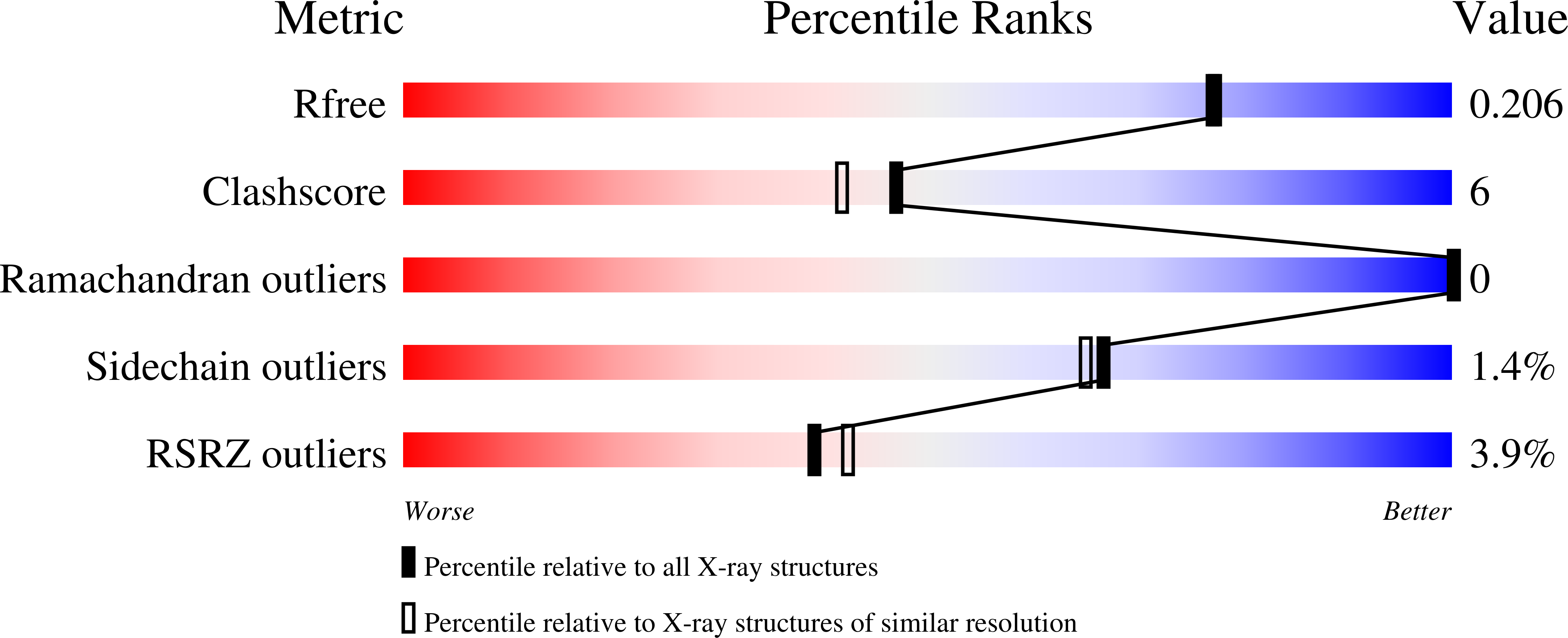
Resolution:
1.90 Å
R-Value Free:
0.22
R-Value Work:
0.19
R-Value Observed:
0.19
Space Group:
P 63 2 2
Macromolecular Entities
Polymer Type:polypeptide(L)
Molecule:HOMOGENTISATE 1,2-DIOXYGENASE
Gene (Uniprot):HGD
Chain IDs:A
Chain Length:471
Number of Molecules:1
Biological Source:Homo sapiens
Modified Residue
Compound ID Chain ID Parent Comp ID Details 2D Image
MSE A MET SELENOMETHIONINE
Primary Citation
Crystal structure of human homogentisate dioxygenase.
Nat.Struct.Biol. 7 542 546 (2000)
PMID: 10876237 DOI: 10.1038/76756

Abstact

Homogentisate dioxygenase (HGO) cleaves the aromatic ring during the metabolic degradation of Phe and Tyr. HGO deficiency causes alkaptonuria (AKU), the first human disease shown to be inherited as a recessive Mendelian trait. Crystal structures of apo-HGO and HGO containing an iron ion have been determined at 1.9 and 2.3 A resolution, respectively. The HGO protomer, which contains a 280-residue N-terminal domain and a 140-residue C-terminal domain, associates as a hexamer arranged as a dimer of trimers. The active site iron ion is coordinated near the interface between subunits in the HGO trimer by a Glu and two His side chains. HGO represents a new structural class of dioxygenases. The largest group of AKU associated missense mutations affect residues located in regions of contact between subunits.

Legend

Protein

Chemical

Disease

Primary Citation of related structures