1EXR image
Deposition Date 2000-05-03
Release Date 2000-09-20
Last Version Date 2024-04-03
Entry Detail
PDB ID:
1EXR
Keywords:
Title:
THE 1.0 ANGSTROM CRYSTAL STRUCTURE OF CA+2 BOUND CALMODULIN
Biological Source:
Source Organism:
Host Organism:
Method Details:
Experimental Method:
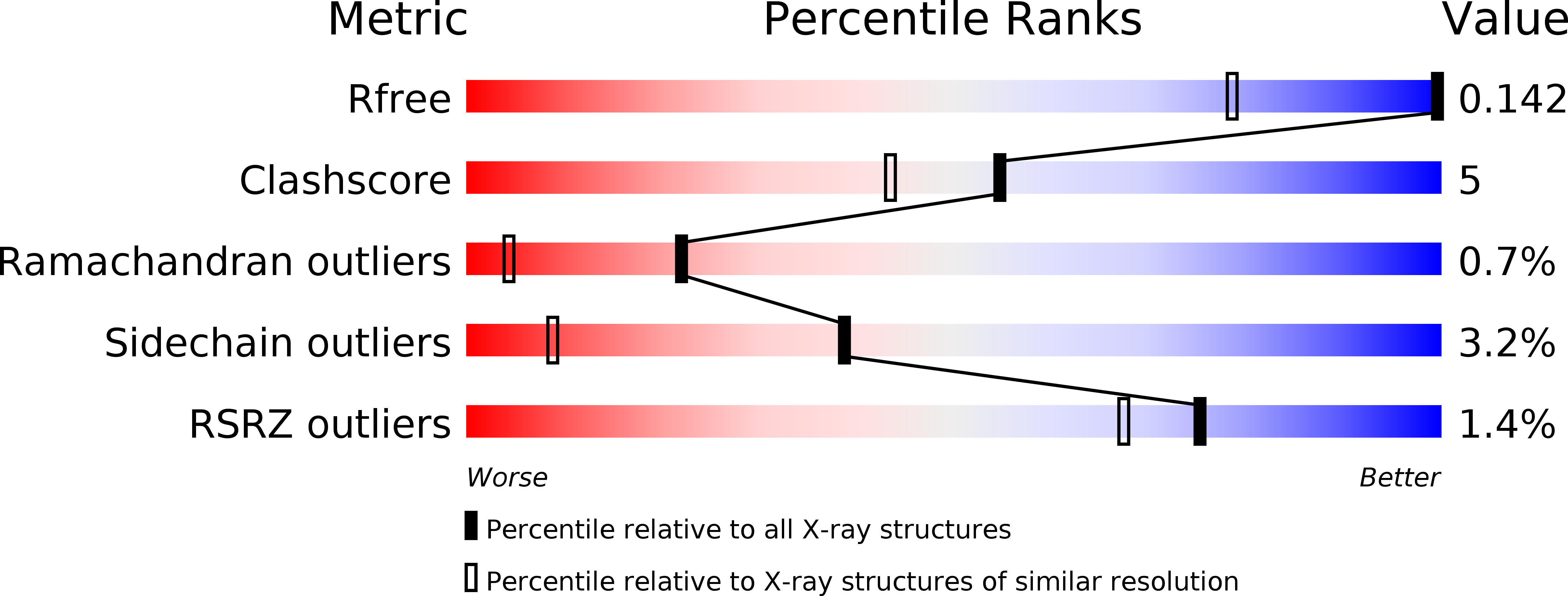
Resolution:
1.00 Å
R-Value Free:
0.16
R-Value Observed:
0.13
Space Group:
P 1
Macromolecular Entities
Polymer Type:polypeptide(L)
Molecule:CALMODULIN
Gene (Uniprot):CAM
Chain IDs:A
Chain Length:148
Number of Molecules:1
Biological Source:Paramecium tetraurelia
Ligand Molecules
Primary Citation
The 1.0 A crystal structure of Ca(2+)-bound calmodulin: an analysis of disorder and implications for functionally relevant plasticity
J.Mol.Biol. 301 1237 1256 (2000)
PMID: 10966818 DOI: 10.1006/jmbi.2000.4029

Abstact

Calmodulin (CaM) is a highly conserved 17 kDa eukaryotic protein that can bind specifically to over 100 protein targets in response to a Ca(2+) signal. Ca(2+)-CaM requires a considerable degree of structural plasticity to accomplish this physiological role; however, the nature and extent of this plasticity remain poorly characterized. Here, we present the 1.0 A crystal structure of Paramecium tetraurelia Ca(2+)-CaM, including 36 discretely disordered residues and a fifth Ca(2+) that mediates a crystal contact. The 36 discretely disordered residues are located primarily in the central helix and the two hydrophobic binding pockets, and reveal correlated side-chain disorder that may assist target-specific deformation of the binding pockets. Evidence of domain displacements and discrete backbone disorder is provided by translation-libration-screw (TLS) analysis and multiconformer models of protein disorder, respectively. In total, the evidence for disorder at every accessible length-scale in Ca(2+)-CaM suggests that the protein occupies a large number of hierarchically arranged conformational substates in the crystalline environment and may sample a quasi-continuous spectrum of conformations in solution. Therefore, we propose that the functionally distinct forms of CaM are less structurally distinct than previously believed, and that the different activities of CaM in response to Ca(2+) may result primarily from Ca(2+)-mediated alterations in the dynamics of the protein.

Legend

Protein

Chemical

Disease

Primary Citation of related structures