1EX9 image
Entry Detail
PDB ID:
1EX9
Keywords:
Title:
CRYSTAL STRUCTURE OF THE PSEUDOMONAS AERUGINOSA LIPASE COMPLEXED WITH RC-(RP,SP)-1,2-DIOCTYLCARBAMOYL-GLYCERO-3-O-OCTYLPHOSPHONATE
Biological Source:
Source Organism:
Host Organism:
PDB Version:
Deposition Date:
2000-05-02
Release Date:
2000-10-18
Method Details:
Experimental Method:
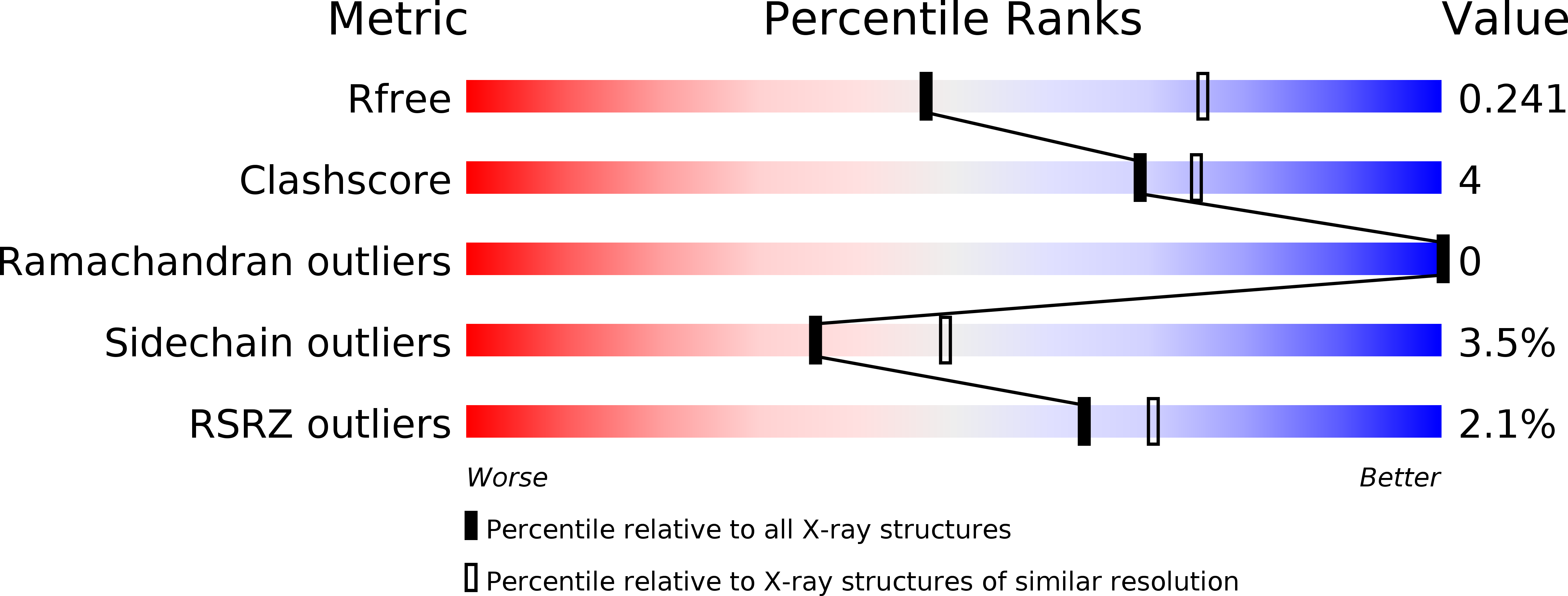
Resolution:
2.54 Å
R-Value Free:
0.24
R-Value Work:
0.19
R-Value Observed:
0.19
Space Group:
P 21 21 21
Macromolecular Entities
Polymer Type:polypeptide(L)
Description:LACTONIZING LIPASE
Chain IDs:A
Chain Length:285
Number of Molecules:1
Biological Source:Pseudomonas aeruginosa
Modified Residue
Compound ID Chain ID Parent Comp ID Details 2D Image
SER A SER COVALENT LINK WITH OCP
Primary Citation
Crystal structure of pseudomonas aeruginosa lipase in the open conformation. The prototype for family I.1 of bacterial lipases.
J.Biol.Chem. 275 31219 31225 (2000)
PMID: 10893416 DOI: 10.1074/jbc.M003903200

Abstact

The x-ray structure of the lipase from Pseudomonas aeruginosa PAO1 has been determined at 2.54 A resolution. It is the first structure of a member of homology family I.1 of bacterial lipases. The structure shows a variant of the alpha/beta hydrolase fold, with Ser(82), Asp(229), and His(251) as the catalytic triad residues. Compared with the "canonical" alpha/beta hydrolase fold, the first two beta-strands and one alpha-helix (alphaE) are not present. The absence of helix alphaE allows the formation of a stabilizing intramolecular disulfide bridge. The loop containing His(251) is stabilized by an octahedrally coordinated calcium ion. On top of the active site a lid subdomain is in an open conformation, making the catalytic cleft accessible from the solvent region. A triacylglycerol analogue is covalently bound to Ser(82) in the active site, demonstrating the position of the oxyanion hole and of the three pockets that accommodate the sn-1, sn-2, and sn-3 fatty acid chains. The inhibited enzyme can be thought to mimic the structure of the tetrahedral intermediate that occurs during the acylation step of the reaction. Analysis of the binding mode of the inhibitor suggests that the size of the acyl pocket and the size and interactions of the sn-2 binding pocket are the predominant determinants of the regio- and enantio-preference of the enzyme.

Legend

Protein

Chemical

Disease

Primary Citation of related structures