
Deposition Date
2000-04-26
Release Date
2000-12-11
Last Version Date
2024-02-07
Entry Detail
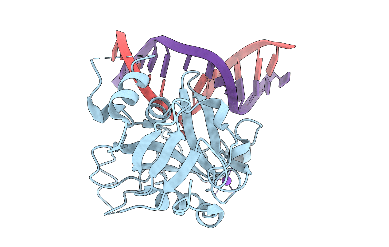
PDB ID:
1EWN
Keywords:
Title:
CRYSTAL STRUCTURE OF THE HUMAN AAG DNA REPAIR GLYCOSYLASE COMPLEXED WITH 1,N6-ETHENOADENINE-DNA
Biological Source:
Source Organism(s):
Homo sapiens (Taxon ID: 9606)
Expression System(s):
Method Details:
Experimental Method:
Resolution:
2.10 Å
R-Value Free:
0.25
R-Value Work:
0.23
Space Group:
P 21 21 21
