1EWK image
Deposition Date 2000-04-26
Release Date 2000-12-18
Last Version Date 2024-10-16
Entry Detail
PDB ID:
1EWK
Title:
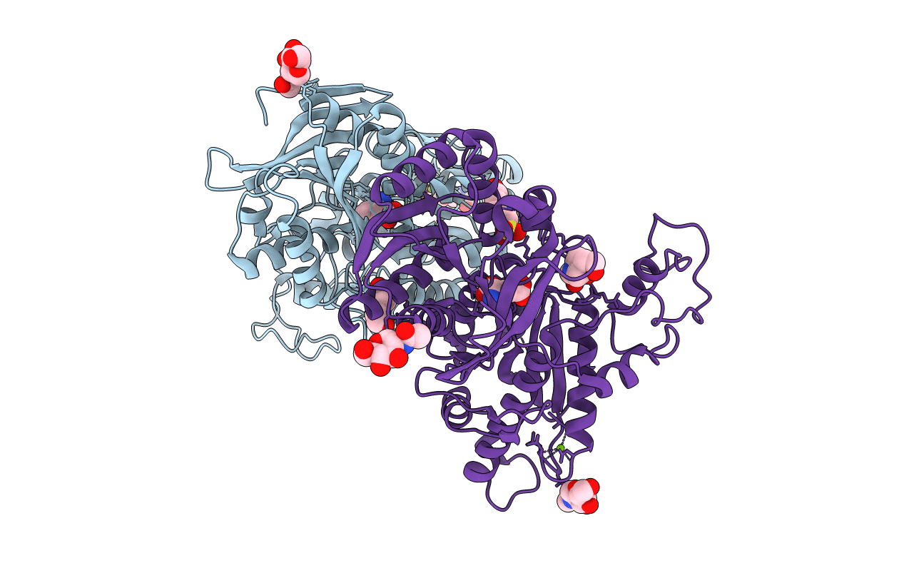
CRYSTAL STRUCTURE OF METABOTROPIC GLUTAMATE RECEPTOR SUBTYPE 1 COMPLEXED WITH GLUTAMATE
Biological Source:
Source Organism:
Host Organism:
Method Details:
Experimental Method:
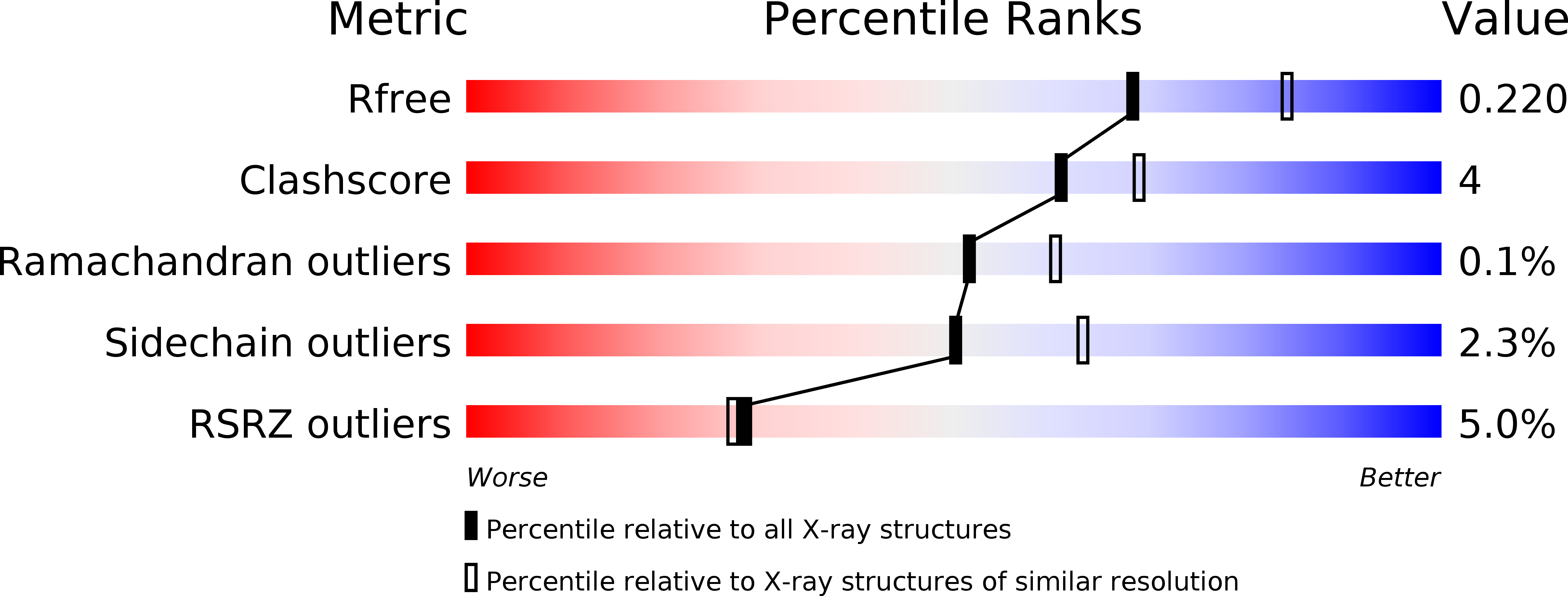
Resolution:
2.20 Å
R-Value Free:
0.22
R-Value Work:
0.19
R-Value Observed:
0.19
Space Group:
P 1 21 1
Macromolecular Entities
Polymer Type:polypeptide(L)
Molecule:METABOTROPIC GLUTAMATE RECEPTOR SUBTYPE 1
Gene (Uniprot):Grm1
Chain IDs:A, B
Chain Length:490
Number of Molecules:2
Biological Source:Rattus norvegicus
Modified Residue
Compound ID Chain ID Parent Comp ID Details 2D Image
ASN B ASN GLYCOSYLATION SITE
Primary Citation
Structural basis of glutamate recognition by a dimeric metabotropic glutamate receptor.
Nature 407 971 977 (2000)
PMID: 11069170 DOI: 10.1038/35039564

Abstact

The metabotropic glutamate receptors (mGluRs) are key receptors in the modulation of excitatory synaptic transmission in the central nervous system. Here we have determined three different crystal structures of the extracellular ligand-binding region of mGluR1--in a complex with glutamate and in two unliganded forms. They all showed disulphide-linked homodimers, whose 'active' and 'resting' conformations are modulated through the dimeric interface by a packed alpha-helical structure. The bi-lobed protomer architectures flexibly change their domain arrangements to form an 'open' or 'closed' conformation. The structures imply that glutamate binding stabilizes both the 'active' dimer and the 'closed' protomer in dynamic equilibrium. Movements of the four domains in the dimer are likely to affect the separation of the transmembrane and intracellular regions, and thereby activate the receptor. This scheme in the initial receptor activation could be applied generally to G-protein-coupled neurotransmitter receptors that possess extracellular ligand-binding sites.

Legend

Protein

Chemical

Disease

Primary Citation of related structures