Deposition Date
2000-04-21
Release Date
2001-02-22
Last Version Date
2024-02-07
Entry Detail
PDB ID:
1EVZ
Keywords:
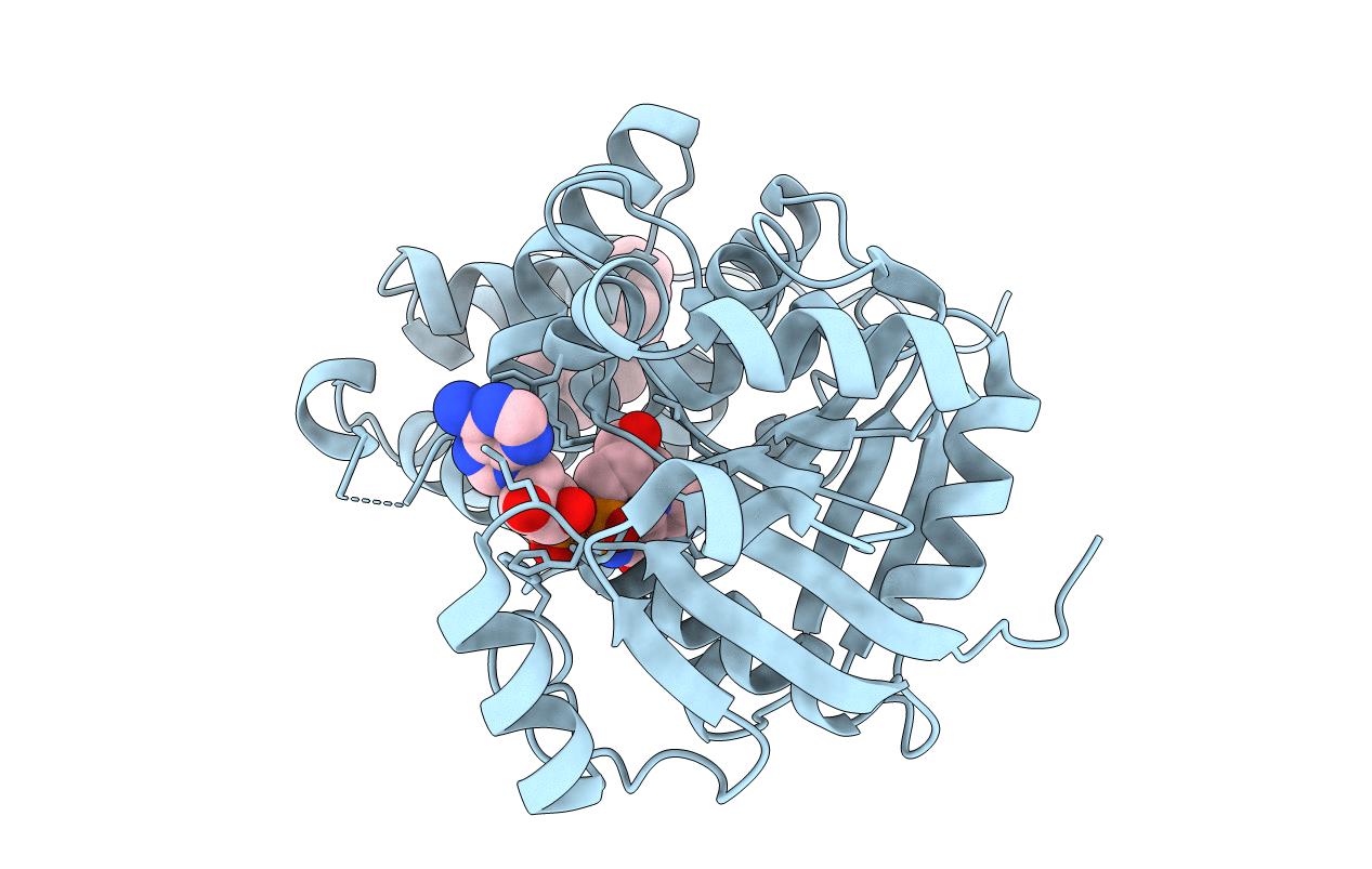
Title:
CRYSTAL STRUCTURE OF LEISHMANIA MEXICANA GLYCEROL-3-PHOSPHATE DEHYDROGENASE IN COMPLEX WITH NAD
Biological Source:
Source Organism(s):
Leishmania mexicana (Taxon ID: 5665)
Expression System(s):
Method Details:
Experimental Method:
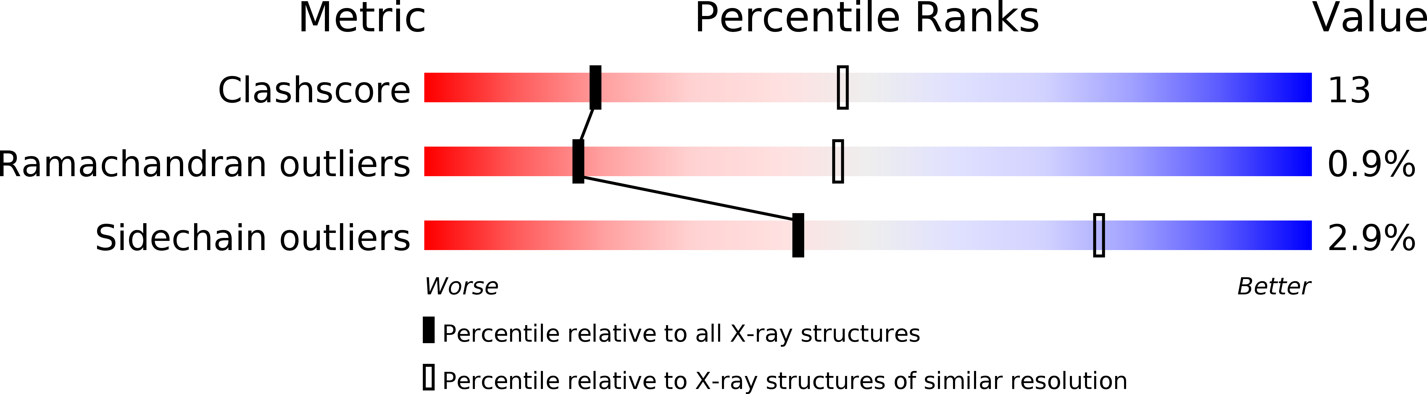
Resolution:
2.80 Å
R-Value Free:
0.25
R-Value Work:
0.18
R-Value Observed:
0.18
Space Group:
P 41 21 2