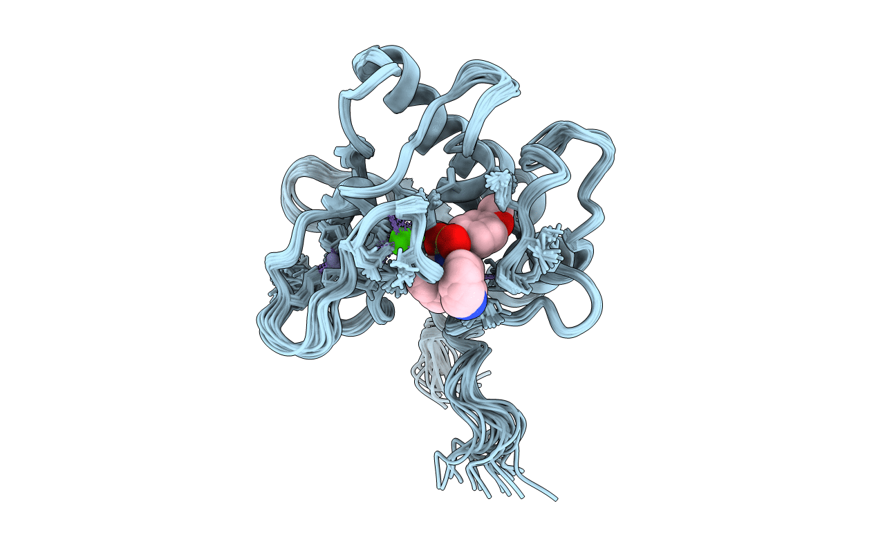
1EUB image
Deposition Date 2000-04-14
Release Date 2001-04-14
Last Version Date 2024-05-22
Entry Detail
PDB ID:
1EUB
Title:
SOLUTION STRUCTURE OF THE CATALYTIC DOMAIN OF HUMAN COLLAGENASE-3 (MMP-13) COMPLEXED TO A POTENT NON-PEPTIDIC SULFONAMIDE INHIBITOR
Biological Source:
Source Organism:
Homo sapiens (Taxon ID: 9606)
Host Organism:
Method Details:
Experimental Method:
Conformers Calculated:
50
Conformers Submitted:
20
Selection Criteria:
BACK CALCULATED DATA AGREE WITH EXPERIMENTAL NOESY SPECTRUM, STRUCTURES WITH ACCEPTABLE COVALENT GEOMETRY,STRUCTURES WITH FAVORABLE NON-BOND ENERGY,TARGET FUNCTION
Macromolecular Entities
Structures with similar UniProt ID
Protein Blast
Polymer Type:polypeptide(L)
Molecule:COLLAGENASE 3
Gene (Uniprot):MMP13
Chain IDs:A
Chain Length:171
Number of Molecules:1
Biological Source:Homo sapiens
Primary Citation
Solution structure of the catalytic domain of human collagenase-3 (MMP-13) complexed to a potent non-peptidic sulfonamide inhibitor: binding comparison with stromelysin-1 and collagenase-1.
J.Mol.Biol. 301 513 524 (2000)
PMID: 10926524 DOI: 10.1006/jmbi.2000.3988

Abstact

The full three-dimensional structure of the catalytic domain of human collagenase-3 (MMP-13) complexed to a potent, sulfonamide hydroxamic acid inhibitor (CGS 27023) has been determined by NMR spectroscopy. The results reveal a core domain for the protein consisting of three alpha-helices and five beta-sheet strands with an overall tertiary fold similar to the catalytic domains of other matrix metalloproteinase family members. The S1' pocket, which is the major site of hydrophobic binding interaction, was found to be a wide cleft spanning the length of the protein and presenting facile opportunity for inhibitor extension deep into the pocket. Comparison with the reported X-ray structure of collagenase-3 showed evidence of flexibility for the loop region flanking the S1' pocket in both NMR and X-ray data. This flexibility was corroborated by NMR dynamics studies. Inhibitor binding placed the methoxy phenyl ring in the S1' pocket with the remainder of the molecule primarily solvent-exposed. The binding mode for this inhibitor was found to be similar with respect to stromelysin-1 and collagenase-1; however, subtle comparative differences in the interactions between inhibitor and enzyme were observed for the three MMPs that were consistent with their respective binding potencies.

Legend

Protein

Chemical

Disease

Primary Citation of related structures