1EU4 image
Deposition Date 2000-04-13
Release Date 2000-04-26
Last Version Date 2024-11-13
Entry Detail
PDB ID:
1EU4
Keywords:
Title:
CRYSTAL STRUCTURE OF THE SUPERANTIGEN SPE-H (ZINC BOUND) FROM STREPTOCOCCUS PYOGENES
Biological Source:
Source Organism:
Host Organism:
Method Details:
Experimental Method:
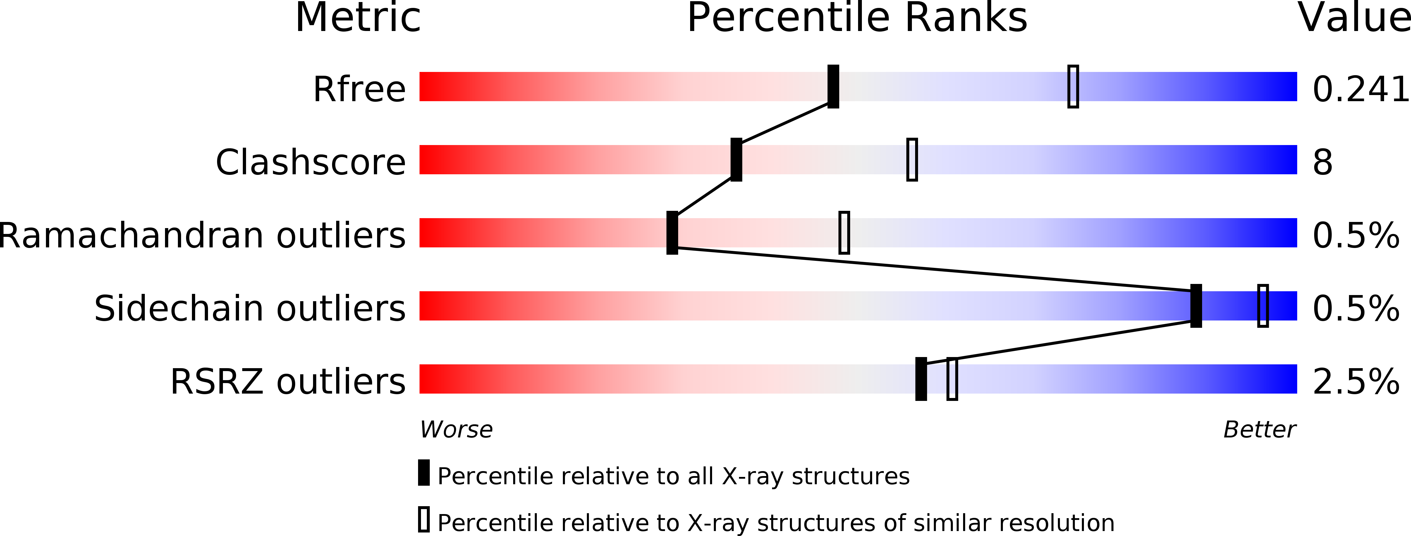
Resolution:
2.50 Å
R-Value Free:
0.25
R-Value Work:
0.20
R-Value Observed:
0.20
Space Group:
P 1 21 1
Macromolecular Entities
Polymer Type:polypeptide(L)
Molecule:SUPERANTIGEN SPE-H
Gene (Uniprot):speH
Chain IDs:A
Chain Length:204
Number of Molecules:1
Biological Source:Streptococcus pyogenes
Ligand Molecules
Primary Citation
Conservation and variation in superantigen structure and activity highlighted by the three-dimensional structures of two new superantigens from Streptococcus pyogenes.
J.Mol.Biol. 299 157 168 (2000)
PMID: 10860729 DOI: 10.1006/jmbi.2000.3725

Abstact

Bacterial superantigens (SAgs) are a structurally related group of protein toxins secreted by Staphylococcus aureus and Streptococcus pyogenes. They are implicated in a range of human pathologies associated with bacterial infection whose symptoms result from SAg-mediated stimulation of a large number (2-20%) of T-cells. At the molecular level, bacterial SAgs bind to major histocompatability class II (MHC-II) molecules and disrupt the normal interaction between MHC-II and T-cell receptors (TCRs). We have determined high-resolution crystal structures of two newly identified streptococcal superantigens, SPE-H and SMEZ-2. Both structures conform to the generic bacterial superantigen folding pattern, comprising an OB-fold N-terminal domain and a beta-grasp C-terminal domain. SPE-H and SMEZ-2 also display very similar zinc-binding sites on the outer concave surfaces of their C-terminal domains. Structural comparisons with other SAgs identify two structural sub-families. Sub-families are related by conserved core residues and demarcated by variable binding surfaces for MHC-II and TCR. SMEZ-2 is most closely related to the streptococcal SAg SPE-C, and together they constitute one structural sub-family. In contrast, SPE-H appears to be a hybrid whose N-terminal domain is most closely related to the SEB sub-family and whose C-terminal domain is most closely related to the SPE-C/SMEZ-2 sub-family. MHC-II binding for both SPE-H and SMEZ-2 is mediated by the zinc ion at their C-terminal face, whereas the generic N-terminal domain MHC-II binding site found on many SAgs appears not to be present. Structural comparisons provide evidence for variations in TCR binding between SPE-H, SMEZ-2 and other members of the SAg family; the extreme potency of SMEZ-2 (active at 10(-15) g ml-1 levels) is likely to be related to its TCR binding properties. The smez gene shows allelic variation that maps onto a considerable proportion of the protein surface. This allelic variation, coupled with the varied binding modes of SAgs to MHC-II and TCR, highlights the pressure on SAgs to avoid host immune defences.

Legend

Protein

Chemical

Disease

Primary Citation of related structures