Deposition Date
1997-06-27
Release Date
1998-07-01
Last Version Date
2024-11-06
Entry Detail
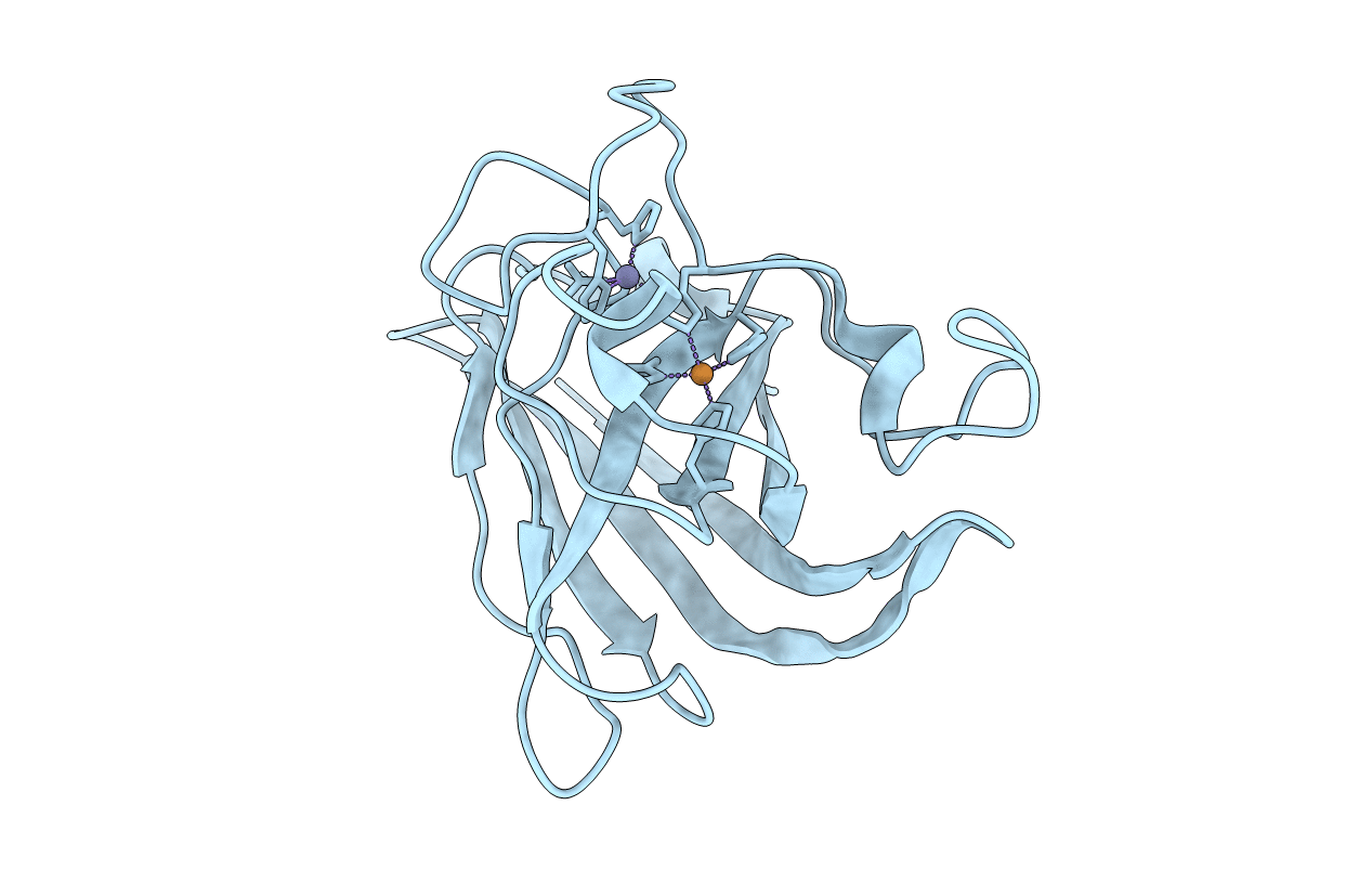
PDB ID:
1ESO
Keywords:
Title:
MONOMERIC CU,ZN SUPEROXIDE DISMUTASE FROM ESCHERICHIA COLI
Biological Source:
Source Organism(s):
Escherichia coli (Taxon ID: 562)
Expression System(s):
Method Details:
Experimental Method:
Resolution:
2.00 Å
R-Value Free:
0.26
R-Value Work:
0.16
Space Group:
P 1 21 1