1ERJ image
Deposition Date 2000-04-06
Release Date 2000-07-26
Last Version Date 2024-05-22
Entry Detail
PDB ID:
1ERJ
Title:
CRYSTAL STRUCTURE OF THE C-TERMINAL WD40 DOMAIN OF TUP1
Biological Source:
Source Organism:
Host Organism:
Method Details:
Experimental Method:
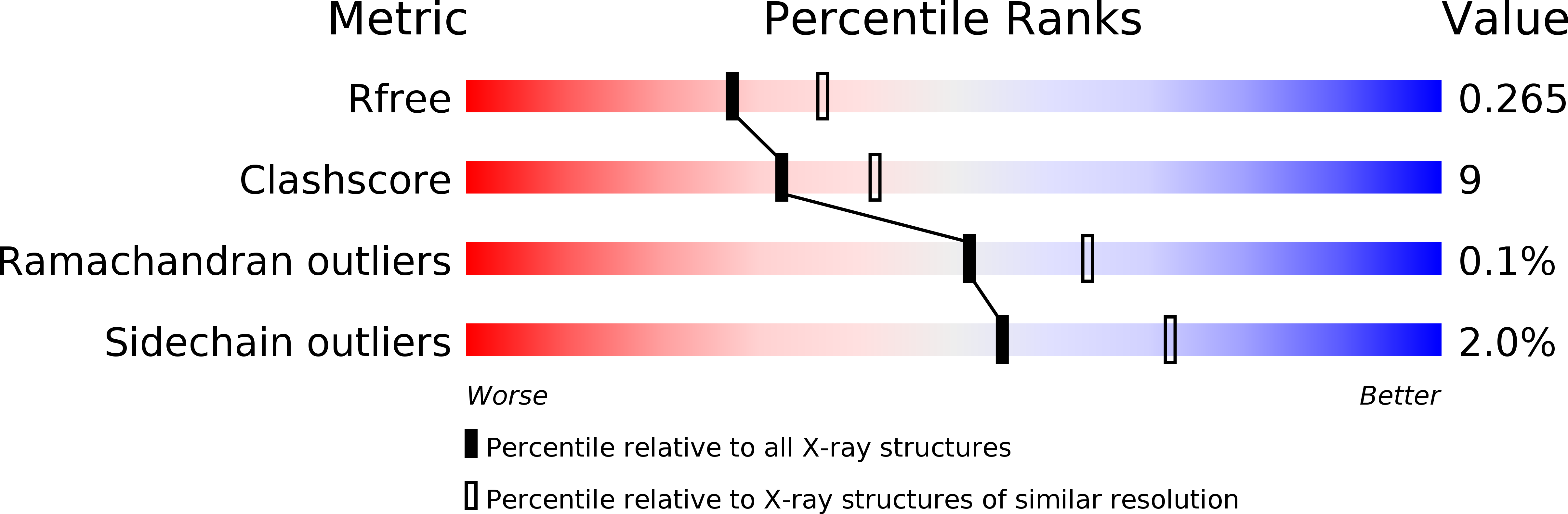
Resolution:
2.30 Å
R-Value Free:
0.26
R-Value Work:
0.22
Space Group:
P 31
Macromolecular Entities
Structures with similar UniProt ID
Protein Blast
Polymer Type:polypeptide(L)
Molecule:TRANSCRIPTIONAL REPRESSOR TUP1
Gene (Uniprot):TUP1
Chain IDs:A, B, C
Chain Length:393
Number of Molecules:3
Biological Source:Saccharomyces cerevisiae
Primary Citation
Structure of the C-terminal domain of Tup1, a corepressor of transcription in yeast.
EMBO J. 19 3016 3027 (2000)
PMID: 10856245 DOI: 10.1093/emboj/19.12.3016

Abstact

The Tup1-Ssn6 corepressor complex regulates the expression of several sets of genes, including genes that specify mating type in the yeast Saccharomyces cerevisiae. Repression of mating-type genes occurs when Tup1-Ssn6 is brought to the DNA by the Matalpha2 DNA-binding protein and assembled upstream of a- and haploid-specific genes. We have determined the 2.3 A X-ray crystal structure of the C-terminal domain of Tup1 (accesion No. 1ERJ), a 43 kDa fragment that contains seven copies of the WD40 sequence motif and binds to the Matalpha2 protein. Moreover, this portion of the protein can partially substitute for full-length Tup1 in bringing about transcriptional repression. The structure reveals a seven-bladed beta propeller with an N-terminal subdomain that is anchored to the side of the propeller and extends the beta sheet of one of the blades. Point mutations in Tup1 that specifically affect the Tup1-Matalpha2 interaction cluster on one surface of the propeller. We identified regions of Tup1 that are conserved among the fungal Tup1 homologs and may be important in protein-protein interactions with additional components of the Tup1-mediated repression pathways.

Legend

Protein

Chemical

Disease

Primary Citation of related structures