1EQR image
Deposition Date 2000-04-06
Release Date 2000-06-29
Last Version Date 2024-04-03
Entry Detail
PDB ID:
1EQR
Keywords:
Title:
CRYSTAL STRUCTURE OF FREE ASPARTYL-TRNA SYNTHETASE FROM ESCHERICHIA COLI
Biological Source:
Source Organism:
Escherichia coli (Taxon ID: 562)
Host Organism:
Method Details:
Experimental Method:
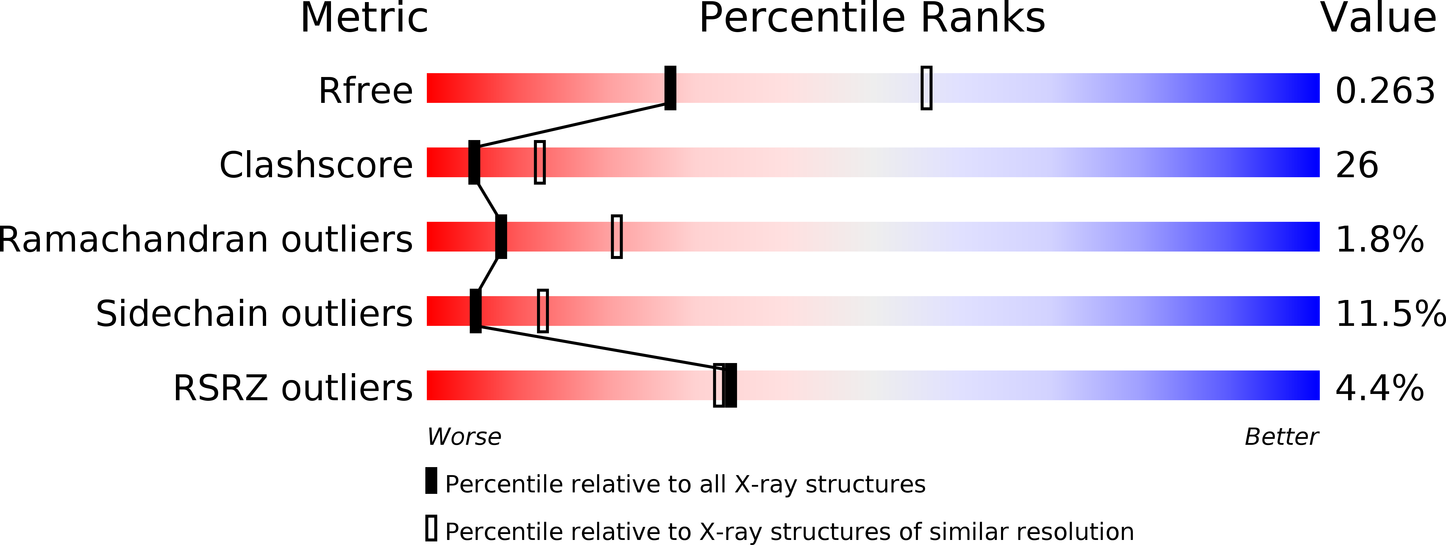
Resolution:
2.70 Å
R-Value Free:
0.26
R-Value Work:
0.19
R-Value Observed:
0.20
Space Group:
C 1 2 1
Macromolecular Entities
Polymer Type:polypeptide(L)
Molecule:ASPARTYL-TRNA SYNTHETASE
Gene (Uniprot):aspS
Chain IDs:A, B, C
Chain Length:590
Number of Molecules:3
Biological Source:Escherichia coli
Ligand Molecules
Primary Citation
Aspartyl tRNA-synthetase from Escherichia coli: flexibility and adaptability to the substrates.
J.Mol.Biol. 299 1157 1164 (2000)
PMID: 10873442 DOI: 10.1006/jmbi.2000.3792

Abstact

The crystal structure of aspartyl-tRNA synthetase from Escherichia coli has been determined to a resolution of 2.7 A. The structure is compared to the same enzyme co-crystallized with tRNA(Asp) and containing aspartyl adenylate or ATP. The asymmetric unit contains three monomers of the enzyme. While most parts of the protein show no significant differences in the three monomers, a few regions cannot be superimposed. Those regions are characterized by a high B-factor, and consist mostly of loops that make contacts with the tRNA in the complexes. The flexibility of the protein is seen at a global level, by the observation of a 10 to 15 degrees rotation of the N-terminal and insertion domains upon tRNA binding, and at the level of the individual amino acid residues, by main-chain and side-chain rearrangements. In contrast to these induced-fit conformational changes, a few residues essential for the tRNA anticodon or aspartyl-adenylate recognition exist in a predefined conformation, ensured by specific interactions within the protein.

Legend

Protein

Chemical

Disease

Primary Citation of related structures