1EPU image
Deposition Date 2000-03-29
Release Date 2000-08-09
Last Version Date 2024-10-30
Entry Detail
PDB ID:
1EPU
Title:
X-RAY crystal structure of neuronal SEC1 from squid
Biological Source:
Source Organism:
Loligo pealei (Taxon ID: 6621)
Host Organism:
Method Details:
Experimental Method:
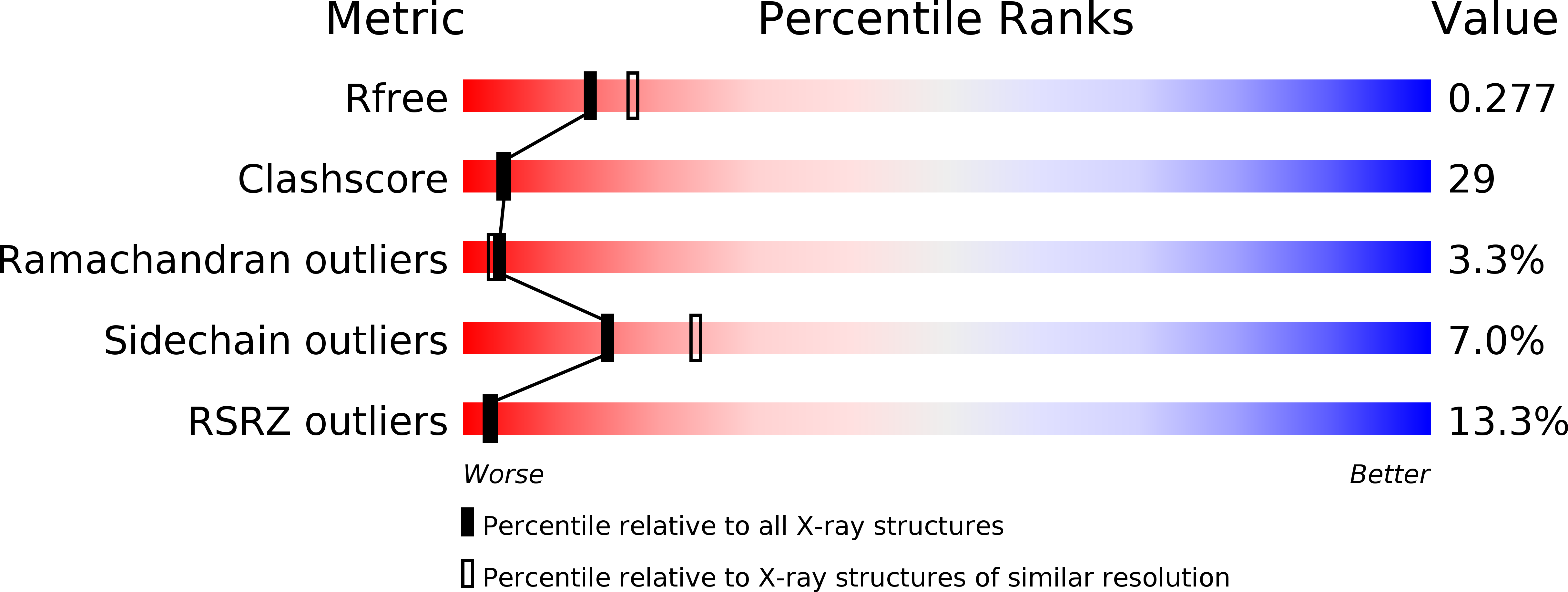
Resolution:
2.40 Å
R-Value Free:
0.27
R-Value Work:
0.24
R-Value Observed:
0.25
Space Group:
P 1 21 1
Macromolecular Entities
Structures with similar UniProt ID
Protein Blast
Polymer Type:polypeptide(L)
Molecule:S-SEC1
Chain IDs:A
Chain Length:591
Number of Molecules:1
Biological Source:Loligo pealei
Modified Residue
Compound ID Chain ID Parent Comp ID Details 2D Image
MSE A MET SELENOMETHIONINE
Primary Citation
The X-ray crystal structure of neuronal Sec1 from squid sheds new light on the role of this protein in exocytosis.
Structure Fold.Des. 8 685 694 (2000)
PMID: 10903948 DOI: 10.1016/S0969-2126(00)00156-8

Abstact

BACKGROUND: Sec1-like molecules have been implicated in a variety of eukaryotic vesicle transport processes including neurotransmitter release by exocytosis. They regulate vesicle transport by binding to a t-SNARE from the syntaxin family. This process is thought to prevent SNARE complex formation, a protein complex required for membrane fusion. Whereas Sec1 molecules are essential for neurotransmitter release and other secretory events, their interaction with syntaxin molecules seems to represent a negative regulatory step in secretion. RESULTS: Here we report the X-ray crystal structure of a neuronal Sec1 homologue from squid, s-Sec1, at 2.4 A resolution. Neuronal s-Sec1 is a modular protein that folds into a V-shaped three-domain assembly. Peptide and mutagenesis studies are discussed with respect to the mechanism of Sec1 regulation. Comparison of the structure of squid s-Sec1 with the previously determined structure of rat neuronal Sec1 (n-Sec1) bound to syntaxin-1a indicates conformational rearrangements in domain III induced by syntaxin binding. CONCLUSIONS: The crystal structure of s-Sec1 provides the molecular scaffold for a number of molecular interactions that have been reported to affect Sec1 function. The structural differences observed between s-Sec1 and the structure of a rat n-Sec1-syntaxin-1a complex suggest that local conformational changes are sufficient to release syntaxin-1a from neuronal Sec1, an active process that is thought to involve additional effector molecule(s).

Legend

Protein

Chemical

Disease

Primary Citation of related structures