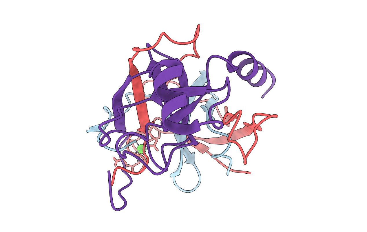
1EPT image
Deposition Date 1994-06-07
Release Date 1995-02-07
Last Version Date 2024-11-20
Entry Detail
PDB ID:
1EPT
Title:
REFINED 1.8 ANGSTROMS RESOLUTION CRYSTAL STRUCTURE OF PORCINE EPSILON-TRYPSIN
Biological Source:
Source Organism:
Sus scrofa (Taxon ID: 9823)
Method Details:
Experimental Method:
Resolution:
1.80 Å
R-Value Work:
0.18
R-Value Observed:
0.18
Space Group:
P 21 21 21
Macromolecular Entities
Structures with similar UniProt ID
Protein Blast
Polymer Type:polypeptide(L)
Molecule:PORCINE E-TRYPSIN
Chain IDs:A
Chain Length:43
Number of Molecules:1
Biological Source:Sus scrofa
Structures with similar UniProt ID
Protein Blast
Polymer Type:polypeptide(L)
Molecule:PORCINE E-TRYPSIN
Chain IDs:B
Chain Length:82
Number of Molecules:1
Biological Source:Sus scrofa
Structures with similar UniProt ID
Protein Blast
Polymer Type:polypeptide(L)
Molecule:PORCINE E-TRYPSIN
Chain IDs:C
Chain Length:98
Number of Molecules:1
Biological Source:Sus scrofa
Ligand Molecules
Primary Citation
Refined 1.8 A resolution crystal structure of the porcine epsilon-trypsin.
Biochim.Biophys.Acta 1209 77 82 (1994)
PMID: 7947985 DOI: 10.1016/0167-4838(94)90139-2

Abstact

Porcine epsilon-trypsin is a three-chain inactivated trypsin from the limited autolysis of porcine beta-trypsin. It is cleaved at positions Lys60-Ser61 and Lys145-Ser146. The crystal structure has been determined by using the molecular replacement method, and refined at 1.8 A resolution. The R-value of final model is 0.184. Comparison with the electron density map of porcine beta-trypsin (PTRY) in complex (BBIT), and with that of native bovine beta-trypsin (HTNA), revealed no obvious changes except at the autolysis positions, and no changes at the active center were observed. The autolysis at positions Lys60-Ser61 and Lys145-Ser146 does not affect the conformation of His-57 in the active center and therefore cannot explain for a loss in porcine epsilon-trypsin activity.

Legend

Protein

Chemical

Disease

Primary Citation of related structures