Deposition Date
2000-03-28
Release Date
2001-12-12
Last Version Date
2024-11-20
Entry Detail
PDB ID:
1EP7
Keywords:
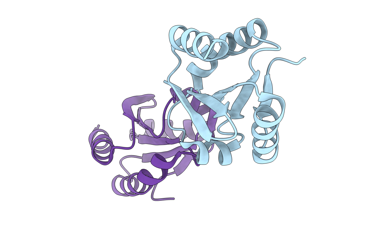
Title:
CRYSTAL STRUCTURE OF WT THIOREDOXIN H FROM CHLAMYDOMONAS REINHARDTII
Biological Source:
Source Organism(s):
Chlamydomonas reinhardtii (Taxon ID: 3055)
Expression System(s):
Method Details:
Experimental Method:
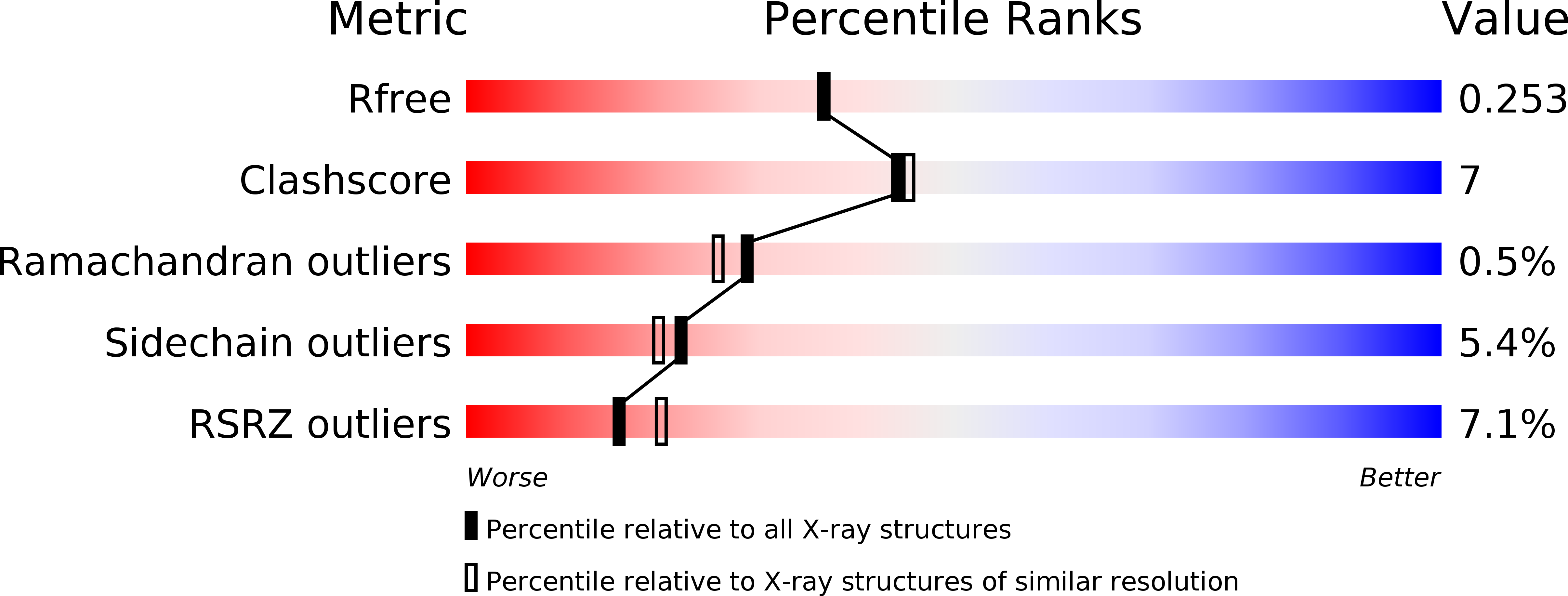
Resolution:
2.10 Å
R-Value Free:
0.25
R-Value Work:
0.20
Space Group:
P 31 2 1