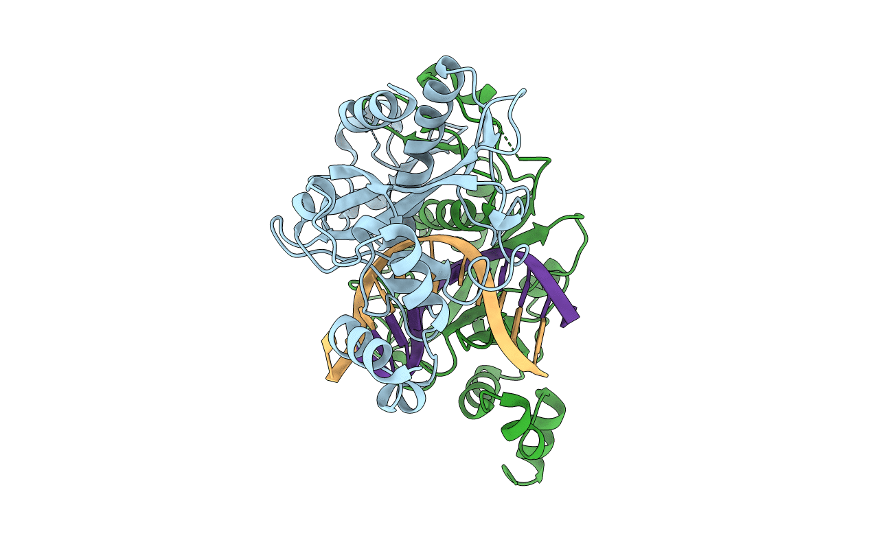
1EOP image
Deposition Date 2000-03-23
Release Date 2000-04-04
Last Version Date 2024-02-07
Entry Detail
PDB ID:
1EOP
Keywords:
Title:
ECORV BOUND TO COGNATE DNA
Biological Source:
Source Organism:
Escherichia coli (Taxon ID: 562)
Host Organism:
Method Details:
Experimental Method:
Resolution:
2.60 Å
R-Value Free:
0.30
R-Value Work:
0.18
R-Value Observed:
0.18
Space Group:
P 41 21 2
Macromolecular Entities
Polymer Type:polypeptide(L)
Molecule:TYPE II RESTRICTION ENZYME ECORV
Gene (Uniprot):ecoRVR
Chain IDs:C (auth: A), D (auth: B)
Chain Length:245
Number of Molecules:2
Biological Source:Escherichia coli
Primary Citation
Crystallographic snapshots along a protein-induced DNA-bending pathway.
Proc.Natl.Acad.Sci.USA 97 5729 5734 (2000)
PMID: 10801972 DOI: 10.1073/pnas.090370797

Abstact

Two new high-resolution cocrystal structures of EcoRV endonuclease bound to DNA show that a large variation in DNA-bending angles is sampled in the ground state binary complex. Together with previous structures, these data reveal a contiguous series of protein conformational states delineating a specific trajectory for the induced-fit pathway. Rotation of the DNA-binding domains, together with movements of two symmetry-related helices binding in the minor groove, causes base unstacking at a key base-pair step and propagates structural changes that assemble the active sites. These structures suggest a complex mechanism for DNA bending that depends on forces generated by interacting protein segments, and on selective neutralization of phosphate charges along the inner face of the bent double helix.

Legend

Protein

Chemical

Disease

Primary Citation of related structures