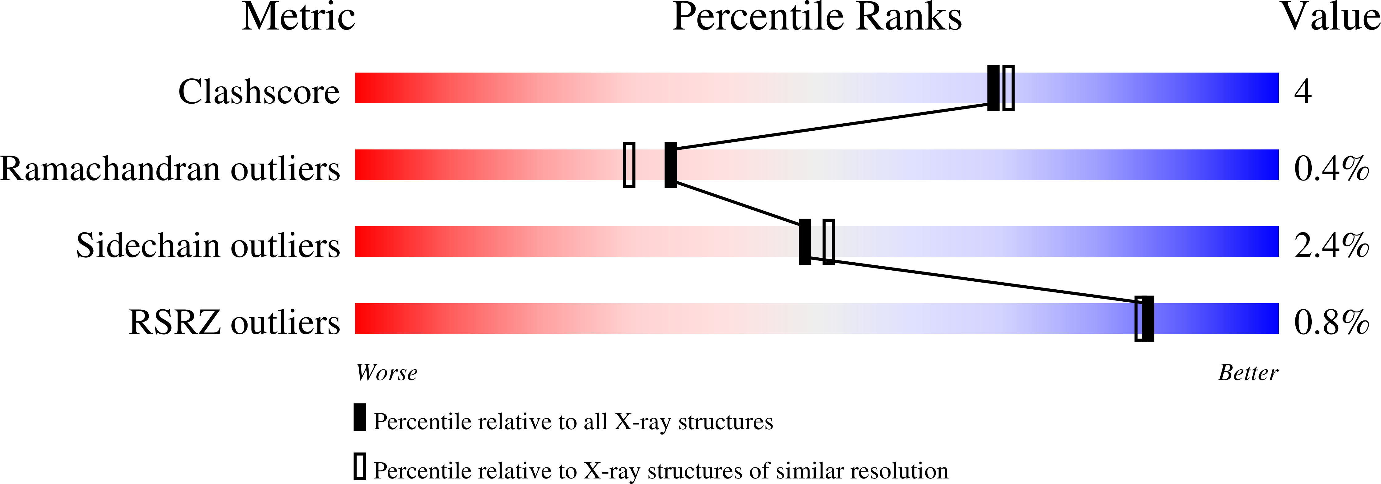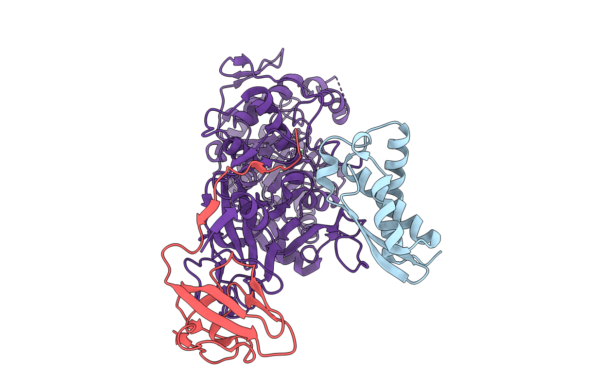
1EJU image
Deposition Date 2000-03-04
Release Date 2000-09-08
Last Version Date 2021-11-03
Entry Detail
PDB ID:
1EJU
Keywords:
Title:
CRYSTAL STRUCTURE OF THE H320N VARIANT OF KLEBSIELLA AEROGENES UREASE
Biological Source:
Source Organism:
Host Organism:
Method Details:
Experimental Method:
Resolution:
2.00 Å
R-Value Work:
0.18
Space Group:
I 21 3
Macromolecular Entities
Polymer Type:polypeptide(L)
Molecule:UREASE GAMMA SUBUNIT
Gene (Uniprot):ureA
Chain IDs:C (auth: A)
Chain Length:100
Number of Molecules:1
Biological Source:Klebsiella aerogenes
Polymer Type:polypeptide(L)
Molecule:UREASE BETA SUBUNIT
Gene (Uniprot):ureB
Chain IDs:B
Chain Length:101
Number of Molecules:1
Biological Source:Klebsiella aerogenes
Polymer Type:polypeptide(L)
Molecule:UREASE ALPHA SUBUNIT
Gene (Uniprot):ureC
Mutations:H320N
Chain IDs:A (auth: C)
Chain Length:567
Number of Molecules:1
Biological Source:Klebsiella aerogenes
Modified Residue
Compound ID Chain ID Parent Comp ID Details 2D Image
KCX A LYS LYSINE NZ-CARBOXYLIC ACID
Ligand Molecules
Primary Citation
Kinetic and structural characterization of urease active site variants.
Biochemistry 39 8575 8584 (2000)
PMID: 10913264 DOI: 10.1021/bi000613o

Abstact

Klebsiella aerogenes urease uses a dinuclear nickel active site to catalyze urea hydrolysis at >10(14)-fold the spontaneous rate. To better define the enzyme mechanism, we examined the kinetics and structures for a suite of site-directed variants involving four residues at the active site: His320, His219, Asp221, and Arg336. Compared to wild-type urease, the H320A, H320N, and H320Q variants exhibit similar approximately 10(-)(5)-fold deficiencies in rates, modest K(m) changes, and disorders in the peptide flap covering their active sites. The pH profiles for these mutant enzymes are anomalous with optima near 6 and shoulders that extend to pH 9. H219A urease exhibits 10(3)-fold increased K(m) over that of native enzyme, whereas the increase is less marked ( approximately 10(2)-fold) in the H219N and H219Q variants that retain hydrogen bonding capability. Structures for these variants show clearly resolved active site water molecules covered by well-ordered peptide flaps. Whereas the D221N variant is only moderately affected compared to wild-type enzyme, D221A urease possesses low activity ( approximately 10(-)(3) that of native enzyme), a small increase in K(m), and a pH 5 optimum. The crystal structure for D221A urease is reminiscent of the His320 variants. The R336Q enzyme has a approximately 10(-)(4)-fold decreased catalytic rate with near-normal pH dependence and an unaffected K(m). Phenylglyoxal inactivates the R336Q variant at over half the rate observed for native enzyme, demonstrating that modification of non-active-site arginines can eliminate activity, perhaps by affecting the peptide flap. Our data favor a mechanism in which His219 helps to polarize the substrate carbonyl group, a metal-bound terminal hydroxide or bridging oxo-dianion attacks urea to form a tetrahedral intermediate, and protonation occurs via the general acid His320 with Asp221 and Arg336 orienting and influencing the acidity of this residue. Furthermore, we conclude that the simple bell-shaped pH dependence of k(cat) and k(cat)/K(m) for the native enzyme masks a more complex underlying pH dependence involving at least four pK(a)s.

Legend

Protein

Chemical

Disease

Primary Citation of related structures