1EJM image
Deposition Date 2000-03-03
Release Date 2001-03-03
Last Version Date 2024-11-20
Entry Detail
PDB ID:
1EJM
Title:
CRYSTAL STRUCTURE OF THE BPTI ALA16LEU MUTANT IN COMPLEX WITH BOVINE TRYPSIN
Biological Source:
Source Organism:
Bos taurus (Taxon ID: 9913)
Host Organism:
Method Details:
Experimental Method:
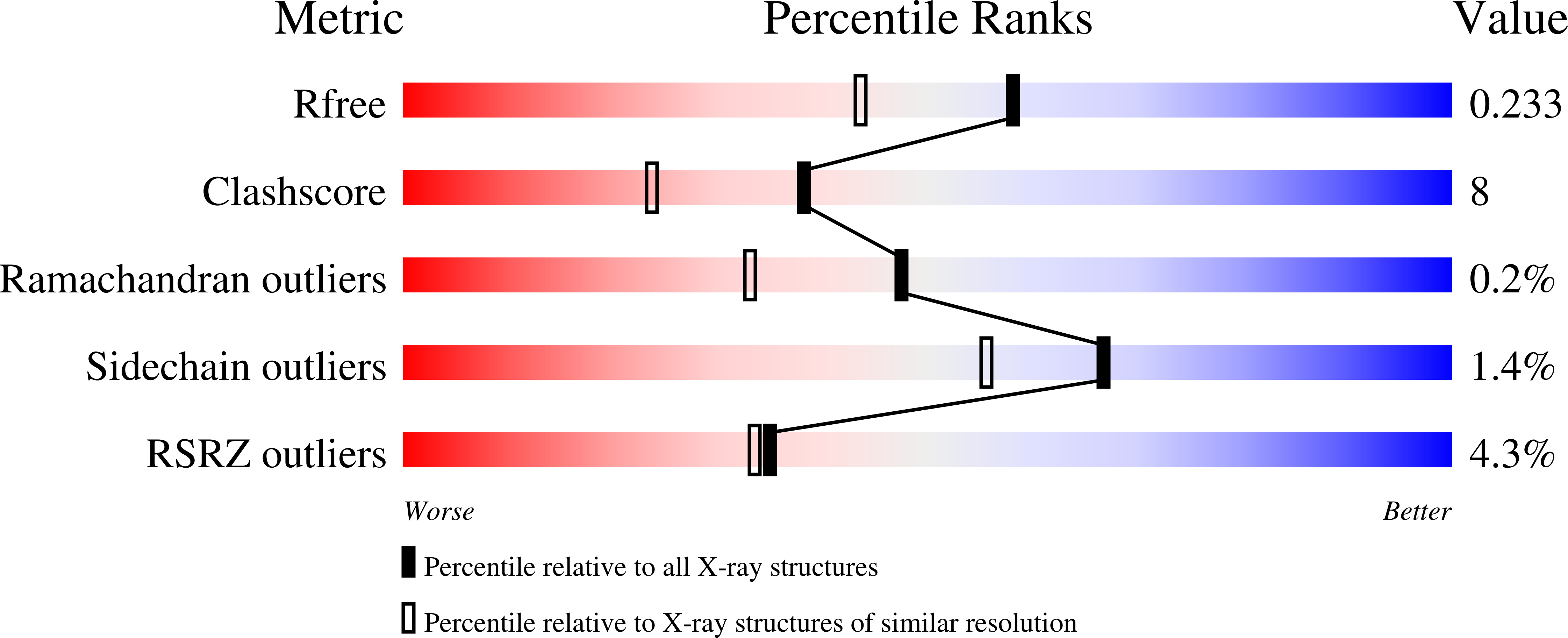
Resolution:
1.85 Å
R-Value Free:
0.23
R-Value Work:
0.21
R-Value Observed:
0.21
Space Group:
I 41 2 2
Macromolecular Entities
Polymer Type:polypeptide(L)
Molecule:BETA-TRYPSIN
Gene (Uniprot):PRSS1
Chain IDs:A, C, E
Chain Length:223
Number of Molecules:3
Biological Source:Bos taurus
Polymer Type:polypeptide(L)
Molecule:PANCREATIC TRYPSIN INHIBITOR
Mutations:K15R, A16L, M52L
Chain IDs:B, D, F
Chain Length:58
Number of Molecules:3
Biological Source:Bos taurus
Ligand Molecules
Primary Citation
Substitutions at the P(1) position in BPTI strongly affect the association energy with serine proteinases.
J.Mol.Biol. 301 205 217 (2000)
PMID: 10926503 DOI: 10.1006/jmbi.2000.3935

Abstact

The role of the S(1) subsite in trypsin, chymotrypsin and plasmin has been examined by measuring the association with seven different mutants of bovine pancreatic trypsin inhibitor (BPTI); the mutants contain Gly, Ala, Ser, Val, Leu, Arg, and Trp at the P(1) position of the reactive site. The effects of substitutions at the P(1) position on the association constants are very large, comprising seven orders of magnitude for trypsin and plasmin, and over five orders for chymotrypsin. All mutants showed a decrease of the association constant to the three proteinases in the same order: Ala>Gly>Ser>Arg>Val>Leu>Trp. Calorimetric and circular dichroism methods showed that none of the P1 substitutions, except the P1-Val mutant, lead to destabilisation of the binding loop conformation. The X-ray structure of the complex formed between bovine beta-trypsin and P(1)-Leu BPTI showed that the P(1)-Leu sterically conflicts with the side-chain of P(3)-Ile, which thereby is forced to rotate approximately 90 degrees. Ile18 (P(3)) in its new orientation, in turn interacts with the Tyr39 side-chain of trypsin. Introduction of a large side-chain at the P1' position apparently leads to a cascade of small alterations of the trypsin-BPTI interface that seem to destabilise the complex by it adopting a less optimized packing and by tilting the BPTI molecule up to 15 degrees compared to the native trypsin-BPTI complex.

Legend

Protein

Chemical

Disease

Primary Citation of related structures