Deposition Date
1998-07-15
Release Date
1999-02-16
Last Version Date
2024-10-09
Entry Detail
PDB ID:
1EIA
Keywords:
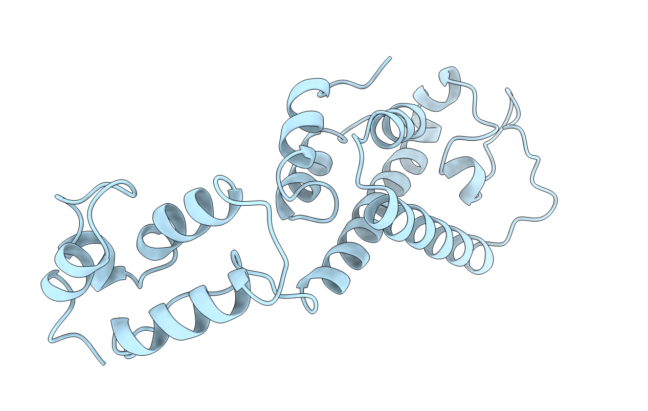
Title:
X-RAY CRYSTAL STRUCTURE OF EQUINE INFECTIOUS ANEMIA VIRUS (EIAV) CAPSID PROTEIN P26
Biological Source:
Source Organism(s):
Equine infectious anemia virus (Taxon ID: 11665)
Expression System(s):
Method Details:
Experimental Method:
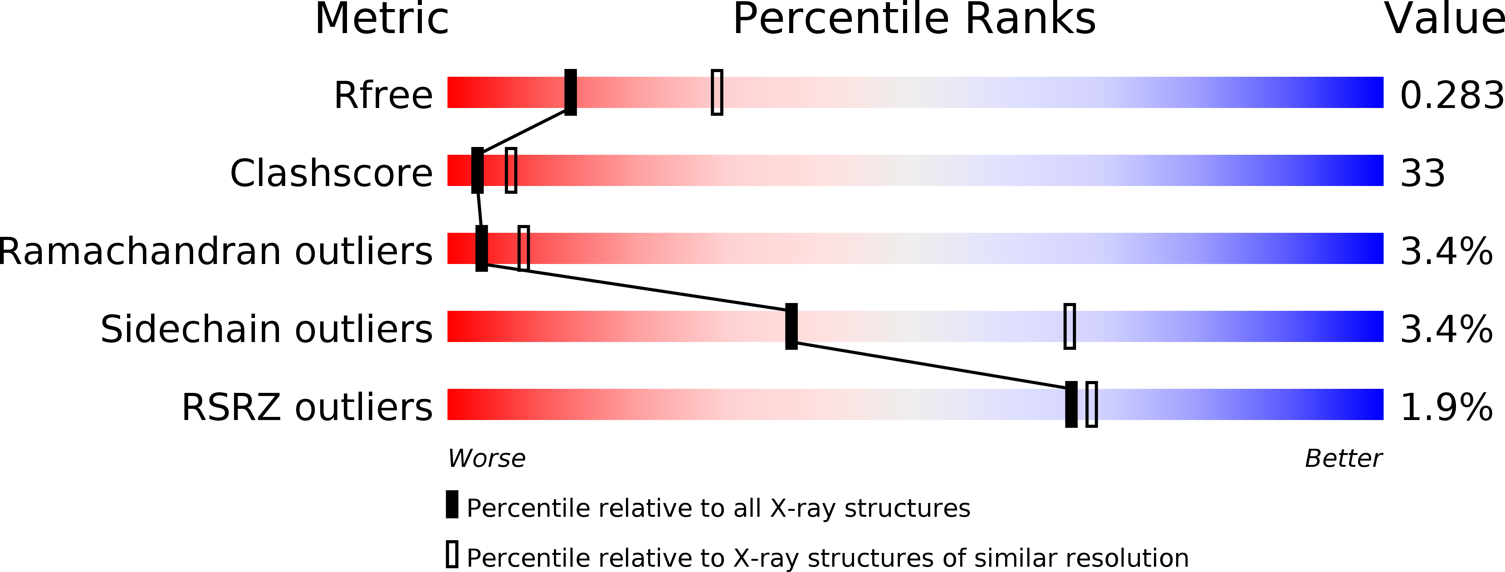
Resolution:
2.70 Å
R-Value Free:
0.27
R-Value Work:
0.22
R-Value Observed:
0.22
Space Group:
I 4 3 2