1EHH image
Deposition Date 2000-02-21
Release Date 2000-04-05
Last Version Date 2024-10-16
Entry Detail
PDB ID:
1EHH
Keywords:
Title:
CRYSTAL STRUCTURE OF URTICA DIOICA AGGLUTININ ISOLECTIN VI COMPLEX WITH TRI-N-ACETYLCHITOTRIOSE
Biological Source:
Source Organism:
Urtica dioica (Taxon ID: 3501)
Method Details:
Experimental Method:
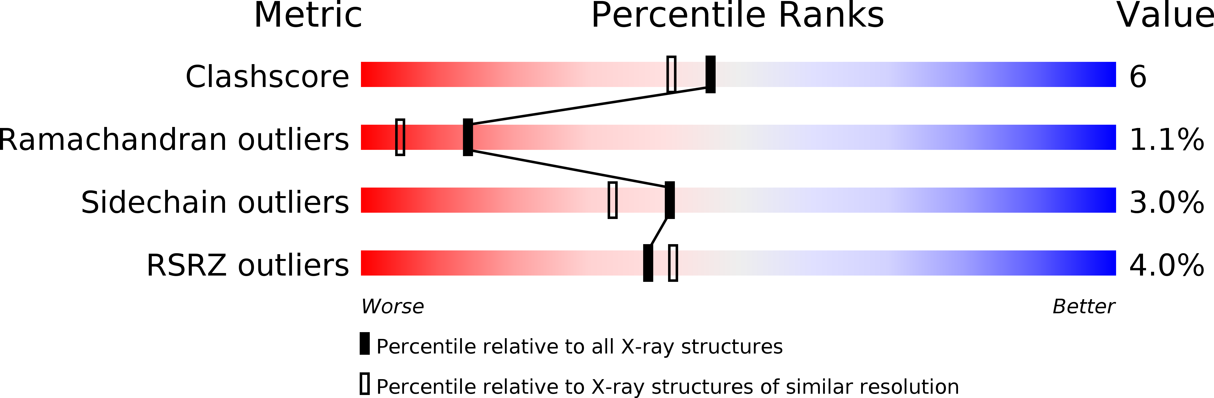
Resolution:
1.90 Å
R-Value Free:
0.25
R-Value Work:
0.18
Space Group:
P 1 21 1
Macromolecular Entities
Polymer Type:polypeptide(L)
Molecule:AGGLUTININ ISOLECTIN VI
Chain IDs:A, B
Chain Length:89
Number of Molecules:2
Biological Source:Urtica dioica
Modified Residue
Compound ID Chain ID Parent Comp ID Details 2D Image
PCA A GLN PYROGLUTAMIC ACID
Peptide-like Molecules
PRD_900017
Primary Citation
Crystal structures of Urtica dioica agglutinin and its complex with tri-N-acetylchitotriose.
J.Mol.Biol. 297 673 681 (2000)
PMID: 10731420 DOI: 10.1006/jmbi.2000.3594

Abstact

Urtica dioica agglutinin is a small plant lectin that binds chitin. We purified the isolectin VI (UDA-VI) and crystal structures of the isolectin and its complex with tri-N-acetylchitotriose (NAG3) were determined by X-ray analysis. The UDA-VI consists of two domains analogous to hevein and the backbone folding of each domain is maintained by four disulfide bridges. The sequence similarity of the two domains is not high (42 %) but their backbone structures are well superimposed except some loop regions. The chitin binding sites are located on the molecular surface at both ends of the dumbbell-shape molecule. The crystal of the NAG3 complex contains two independent molecules forming a protein-sugar 2:2 complex. One NAG3 molecule is sandwiched between two independent UDA-VI molecules and the other sugar molecule is also sandwiched by one UDA-VI molecule and symmetry-related another one. The sugar binding site of N-terminal domain consists of three subsites accommodating NAG3 while two NAG residues are bound to the C-terminal domain. In each sugar-binding site, three aromatic amino acid residues and one serine residue participate to the NAG3 binding. The sugar rings bound to two subsites are stacked to the side-chain groups of tryptophan or histidine and a tyrosine residue is in face-to-face contact with an acetylamino group, to which the hydroxyl group of a serine residue is hydrogen-bonded. The third subsite of the N-terminal domain binds a NAG moiety with hydrogen bonds. The results suggest that the triad of aromatic amino acid residues is intrinsic in sugar binding of hevein-like domains.

Legend

Protein

Chemical

Disease

Primary Citation of related structures