1EG4 image
Deposition Date 2000-02-11
Release Date 2000-08-23
Last Version Date 2024-02-07
Entry Detail
PDB ID:
1EG4
Title:
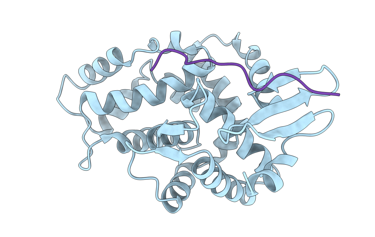
STRUCTURE OF A DYSTROPHIN WW DOMAIN FRAGMENT IN COMPLEX WITH A BETA-DYSTROGLYCAN PEPTIDE
Biological Source:
Source Organism:
Homo sapiens (Taxon ID: 9606)
(Taxon ID: )
Host Organism:
Method Details:
Experimental Method:
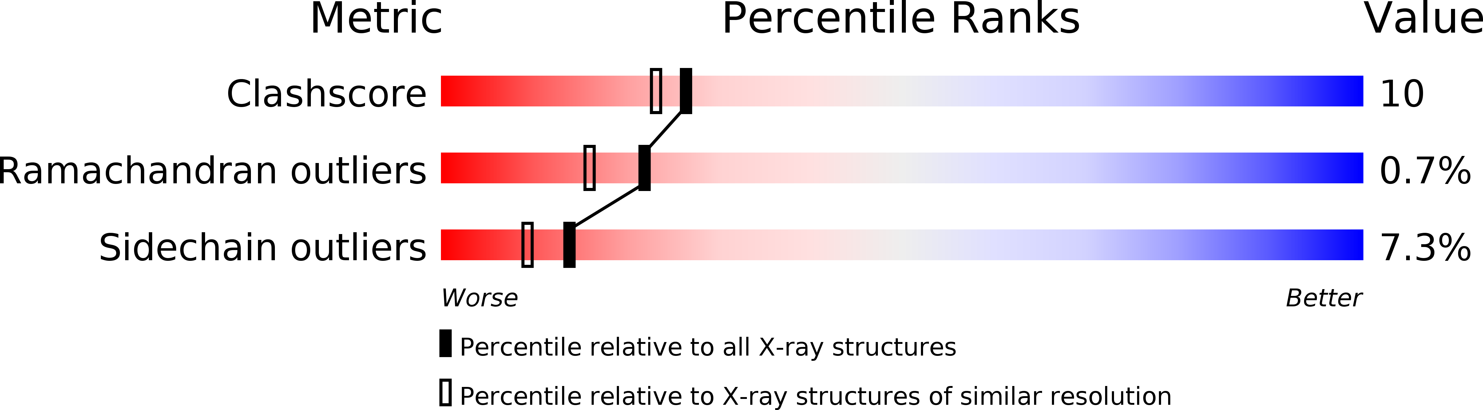
Resolution:
2.00 Å
R-Value Free:
0.24
R-Value Work:
0.19
Space Group:
P 21 21 21
Macromolecular Entities
Polymer Type:polypeptide(L)
Molecule:DYSTROPHIN
Gene (Uniprot):DMD
Chain IDs:B (auth: A)
Chain Length:261
Number of Molecules:1
Biological Source:Homo sapiens
Polymer Type:polypeptide(L)
Molecule:BETA-DYSTROGLYCAN
Gene (Uniprot):DAG1
Chain IDs:A (auth: P)
Chain Length:15
Number of Molecules:1
Biological Source:
Primary Citation
Structure of a WW domain containing fragment of dystrophin in complex with beta-dystroglycan.
Nat.Struct.Biol. 7 634 638 (2000)
PMID: 10932245 DOI: 10.1038/77923

Abstact

Dystrophin and beta-dystroglycan are components of the dystrophin-glycoprotein complex (DGC), a multimolecular assembly that spans the cell membrane and links the actin cytoskeleton to the extracellular basal lamina. Defects in the dystrophin gene are the cause of Duchenne and Becker muscular dystrophies. The C-terminal region of dystrophin binds the cytoplasmic tail of beta-dystroglycan, in part through the interaction of its WW domain with a proline-rich motif in the tail of beta-dystroglycan. Here we report the crystal structure of this portion of dystrophin in complex with the proline-rich binding site in beta-dystroglycan. The structure shows that the dystrophin WW domain is embedded in an adjacent helical region that contains two EF-hand-like domains. The beta-dystroglycan peptide binds a composite surface formed by the WW domain and one of these EF-hands. Additionally, the structure reveals striking similarities in the mechanisms of proline recognition employed by WW domains and SH3 domains.

Legend

Protein

Chemical

Disease

Primary Citation of related structures