
Deposition Date
1996-10-16
Release Date
1997-12-03
Last Version Date
2024-02-07
Entry Detail
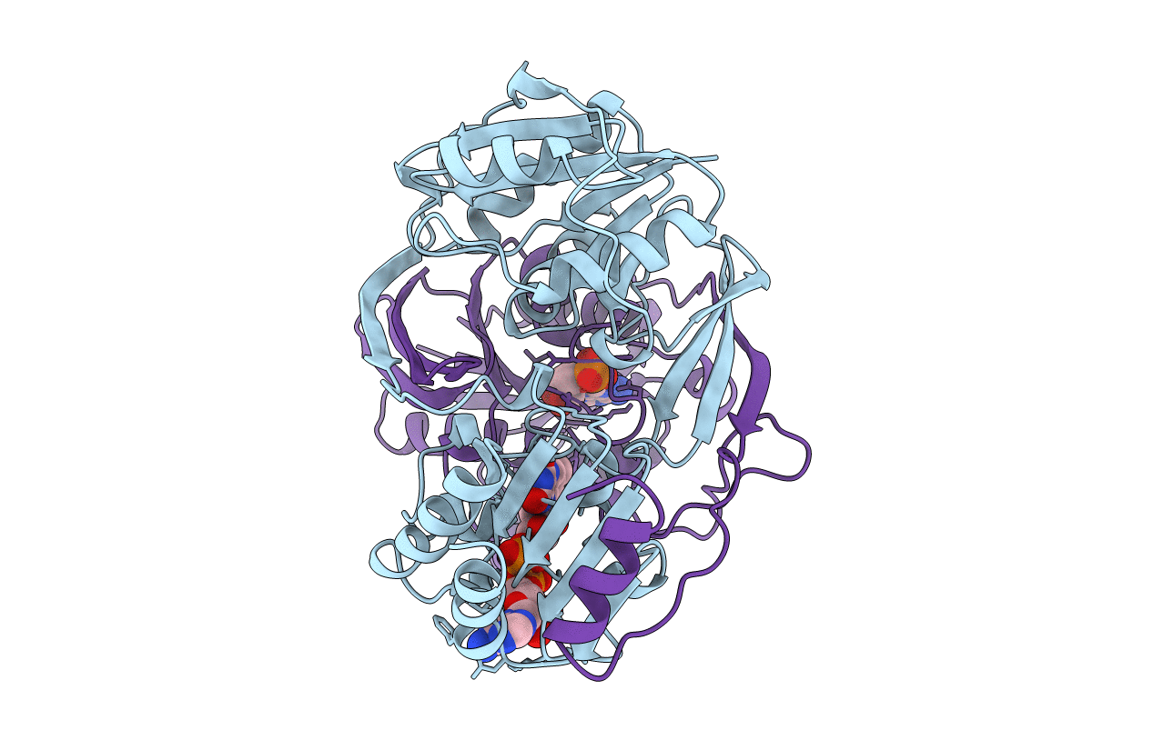
PDB ID:
1EFV
Keywords:
Title:
THREE-DIMENSIONAL STRUCTURE OF HUMAN ELECTRON TRANSFER FLAVOPROTEIN TO 2.1 A RESOLUTION
Biological Source:
Source Organism(s):
Homo sapiens (Taxon ID: 9606)
Expression System(s):
Method Details:
Experimental Method:
Resolution:
2.10 Å
R-Value Free:
0.22
R-Value Work:
0.17
R-Value Observed:
0.17
Space Group:
P 1 21 1
