Deposition Date
2000-02-04
Release Date
2000-05-10
Last Version Date
2024-11-06
Entry Detail
PDB ID:
1EF1
Keywords:
Title:
CRYSTAL STRUCTURE OF THE MOESIN FERM DOMAIN/TAIL DOMAIN COMPLEX
Biological Source:
Source Organism(s):
Homo sapiens (Taxon ID: 9606)
Expression System(s):
Method Details:
Experimental Method:
Resolution:
1.90 Å
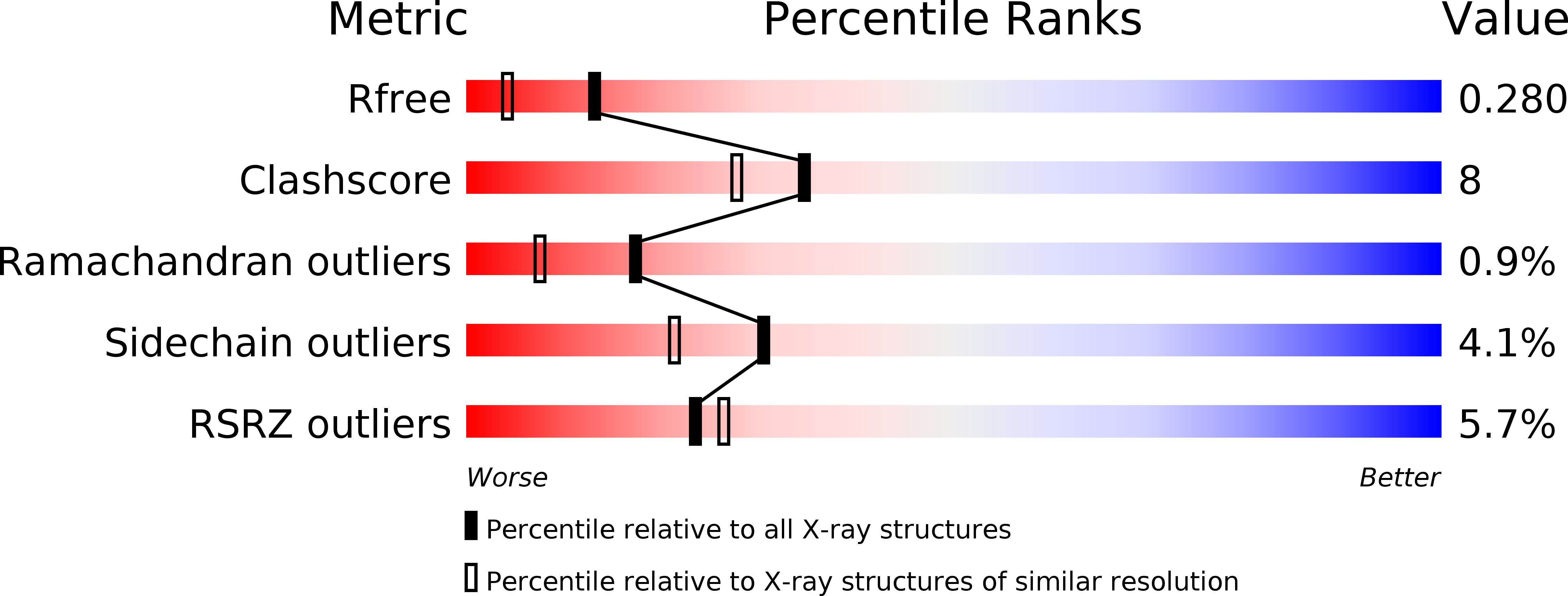
R-Value Free:
0.28
R-Value Work:
0.22
Space Group:
P 2 2 21