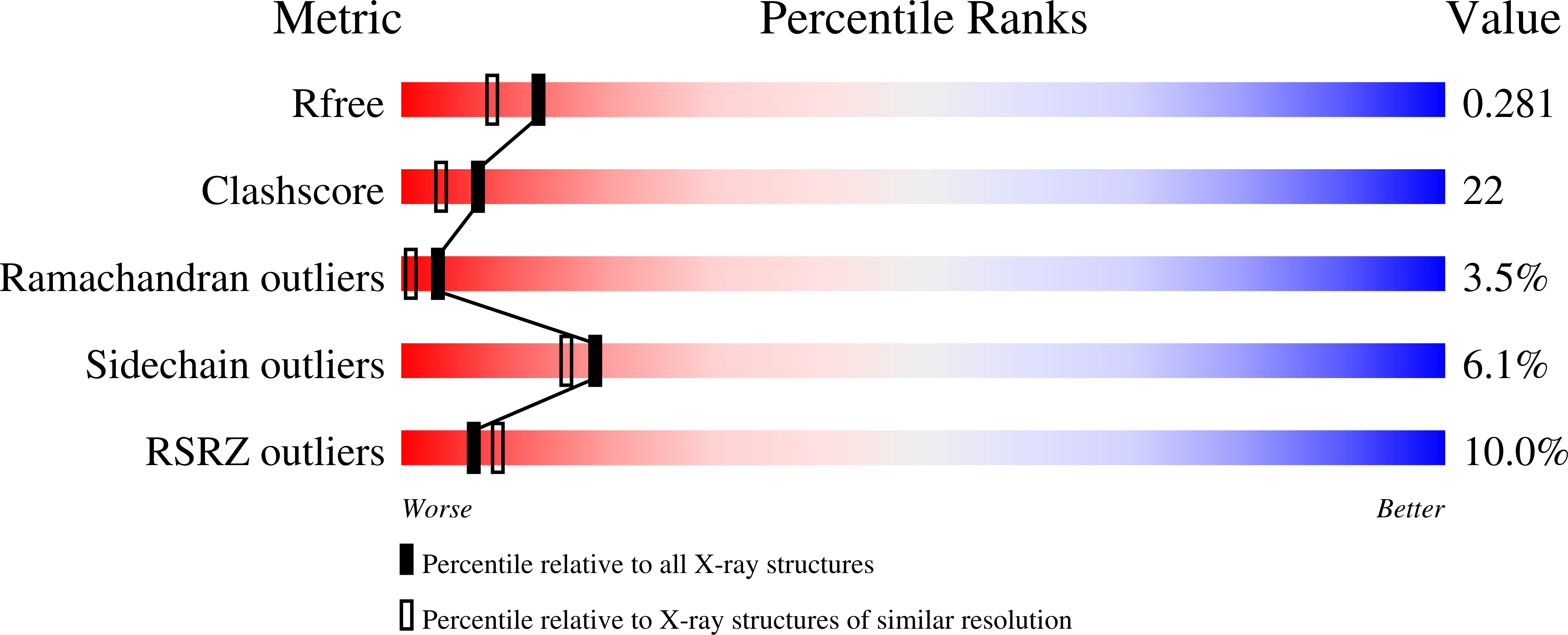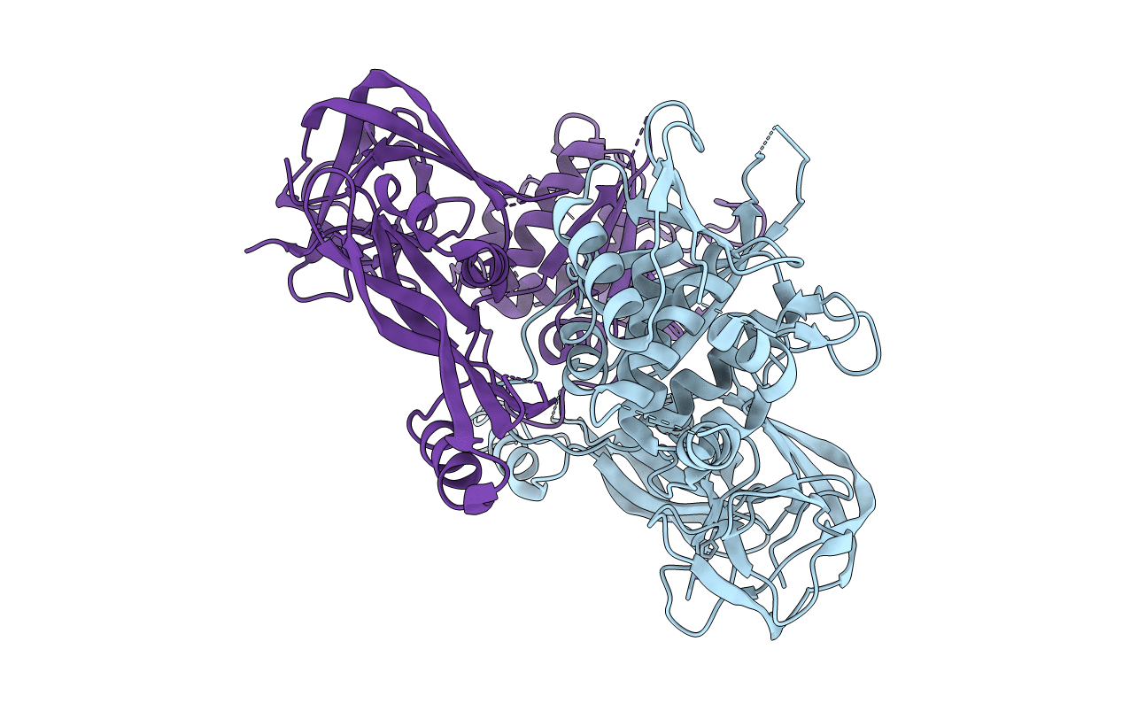
1EF0 image
Deposition Date 2000-02-04
Release Date 2000-06-07
Last Version Date 2024-02-07
Entry Detail
PDB ID:
1EF0
Keywords:
Title:
CRYSTAL STRUCTURE OF PI-SCEI MINIPRECURSOR
Biological Source:
Source Organism:
Host Organism:
Method Details:
Experimental Method:
Resolution:
2.10 Å
R-Value Free:
0.28
R-Value Work:
0.23
R-Value Observed:
0.23
Space Group:
P 1 21 1
Macromolecular Entities
Polymer Type:polypeptide(L)
Molecule:PI-SCEI ENDONUCLEASE
Chain IDs:A, B
Chain Length:462
Number of Molecules:2
Biological Source:Saccharomyces cerevisiae
Ligand Molecules
Primary Citation
Structural insights into the protein splicing mechanism of PI-SceI.
J.Biol.Chem. 275 16408 16413 (2000)
PMID: 10828056 DOI: 10.1074/jbc.275.22.16408

Abstact

PI-SceI is a member of a class of proteins (inteins) that excise themselves from a precursor protein and in the process ligate the flanking protein sequences (exteins). We report here the 2.1-A resolution crystal structure of a PI-SceI miniprecursor (VMA29) containing 10 N-terminal extein residues and 4 C-terminal extein residues. Mutations at the N- and C-terminal splicing junctions, blocking in vivo protein splicing, allowed the miniprecursor to be purified and crystallized. The structure reveals both the N- and C-terminal scissile peptide bonds to be in distorted trans conformations (tau approximately 100 degrees ). Modeling of the wild-type PI-SceI based on the VMA29 structure indicates a large conformational change (movement of >9 A) must occur to allow transesterification to be completed. A zinc atom was discovered at the C-terminal splicing junction. Residues Cys(455), His(453), and Glu(80) along with a water molecule (Wat(53)) chelate the zinc atom. The crystal structure of VMA29 has captured the intein in its pre-spliced state.

Legend

Protein

Chemical

Disease

Primary Citation of related structures