1EEX image
Deposition Date 2000-02-04
Release Date 2001-02-07
Last Version Date 2024-03-13
Entry Detail
PDB ID:
1EEX
Keywords:
Title:
CRYSTAL STRUCTURE OF THE DIOL DEHYDRATASE-ADENINYLPENTYLCOBALAMIN COMPLEX FROM KLEBSIELLA OXYTOCA
Biological Source:
Source Organism:
Host Organism:
Method Details:
Experimental Method:
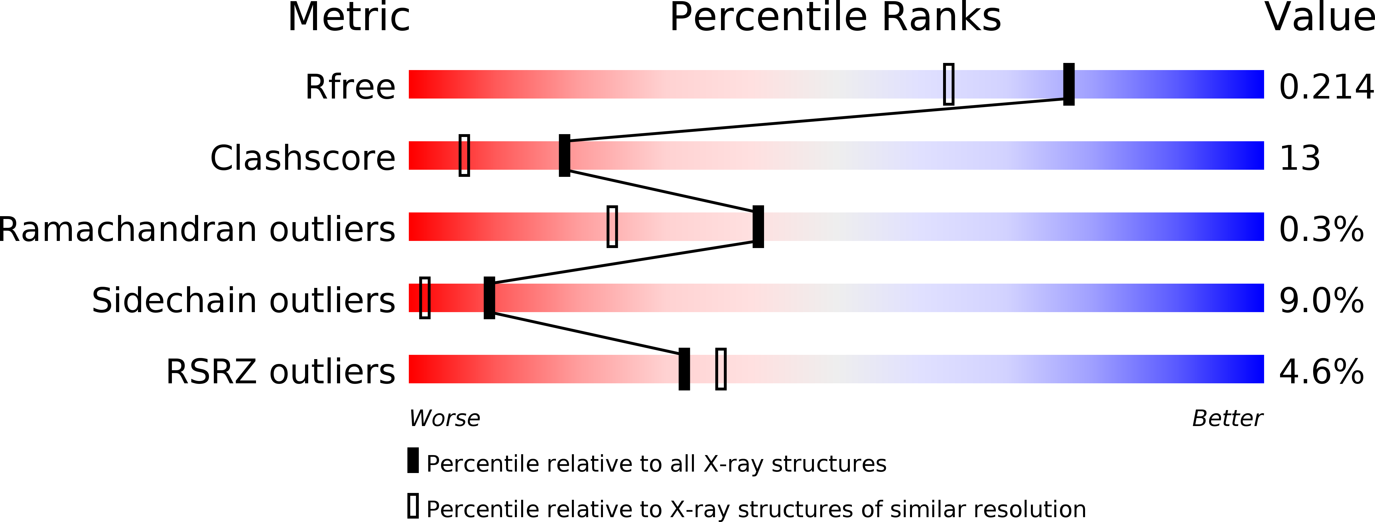
Resolution:
1.70 Å
R-Value Free:
0.22
R-Value Work:
0.16
R-Value Observed:
0.16
Space Group:
P 21 21 21
Macromolecular Entities
Polymer Type:polypeptide(L)
Molecule:PROPANEDIOL DEHYDRATASE
Gene (Uniprot):pddA
Chain IDs:A, D (auth: L)
Chain Length:554
Number of Molecules:2
Biological Source:Klebsiella oxytoca
Polymer Type:polypeptide(L)
Molecule:PROPANEDIOL DEHYDRATASE
Gene (Uniprot):pddB
Chain IDs:B, E
Chain Length:224
Number of Molecules:2
Biological Source:Klebsiella oxytoca
Polymer Type:polypeptide(L)
Molecule:PROPANEDIOL DEHYDRATASE
Gene (Uniprot):pddC
Chain IDs:C (auth: G), F (auth: M)
Chain Length:173
Number of Molecules:2
Biological Source:Klebsiella oxytoca
Primary Citation
How a protein generates a catalytic radical from coenzyme B(12): X-ray structure of a diol-dehydratase-adeninylpentylcobalamin complex.
Structure Fold.Des. 8 775 788 (2000)
PMID: 10903944 DOI: 10.1016/S0969-2126(00)00164-7

Abstact

BACKGROUND: Adenosylcobalamin (coenzyme B(12)) serves as a cofactor for enzymatic radical reactions. The adenosyl radical, a catalytic radical in these reactions, is formed by homolysis of the cobalt-carbon bond of the coenzyme, although the mechanism of cleavage of its organometallic bond remains unsolved. RESULTS: We determined the three-dimensional structures of diol dehydratase complexed with adeninylpentylcobalamin and with cyanocobalamin at 1.7 A and 1.9 A resolution, respectively, at cryogenic temperatures. In the adeninylpentylcobalamin complex, the adenine ring is bound parallel to the corrin ring as in the free form and methylmalonyl-CoA-mutase-bound coenzyme, but with the other side facing pyrrole ring C. All of its nitrogen atoms except for N(9) are hydrogen-bonded to mainchain amide oxygen and amide nitrogen atoms, a sidechain hydroxyl group, and a water molecule. As compared with the cyanocobalamin complex, the sidechain of Seralpha224 rotates by 120 degrees to hydrogen bond with N(3) of the adenine ring. CONCLUSIONS: The structure of the adenine-ring-binding site provides a molecular basis for the strict specificity of diol dehydratase for the coenzyme adenosyl group. The superimposition of the structure of the free coenzyme on that of enzyme-bound adeninylpentylcobalamin demonstrated that the tight enzyme-coenzyme interactions at both the cobalamin moiety and adenine ring of the adenosyl group would inevitably lead to cleavage of the cobalt-carbon bond. Rotation of the ribose moiety around the glycosidic linkage makes the 5'-carbon radical accessible to the hydrogen atom of the substrate to be abstracted.

Legend

Protein

Chemical

Disease

Primary Citation of related structures