Deposition Date
2000-01-31
Release Date
2001-01-31
Last Version Date
2024-10-30
Entry Detail
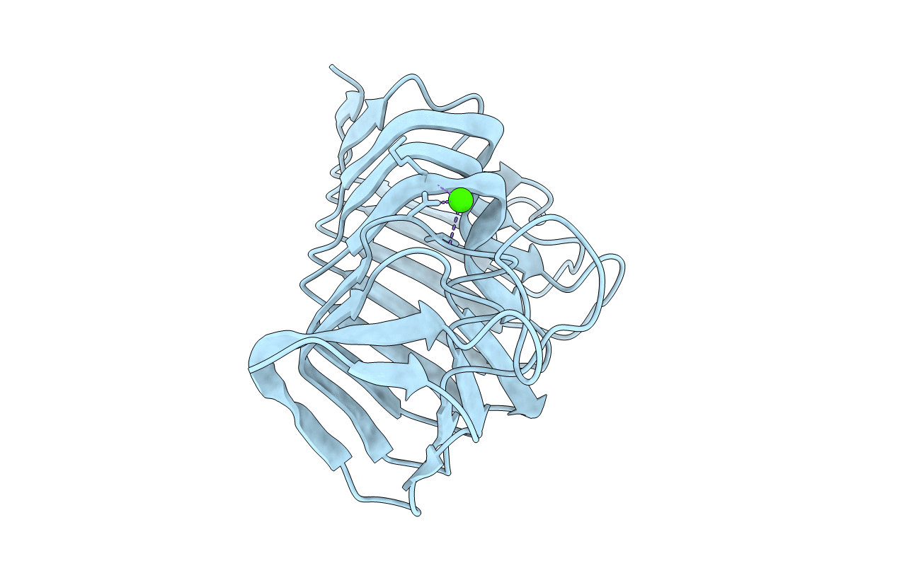
PDB ID:
1EE6
Keywords:
Title:
CRYSTAL STRUCTURE OF PECTATE LYASE FROM BACILLUS SP. STRAIN KSM-P15.
Biological Source:
Source Organism(s):
Bacillus sp. (Taxon ID: 98226)
Method Details:
Experimental Method:
Resolution:
2.30 Å
R-Value Free:
0.23
R-Value Work:
0.18
Space Group:
P 21 21 21