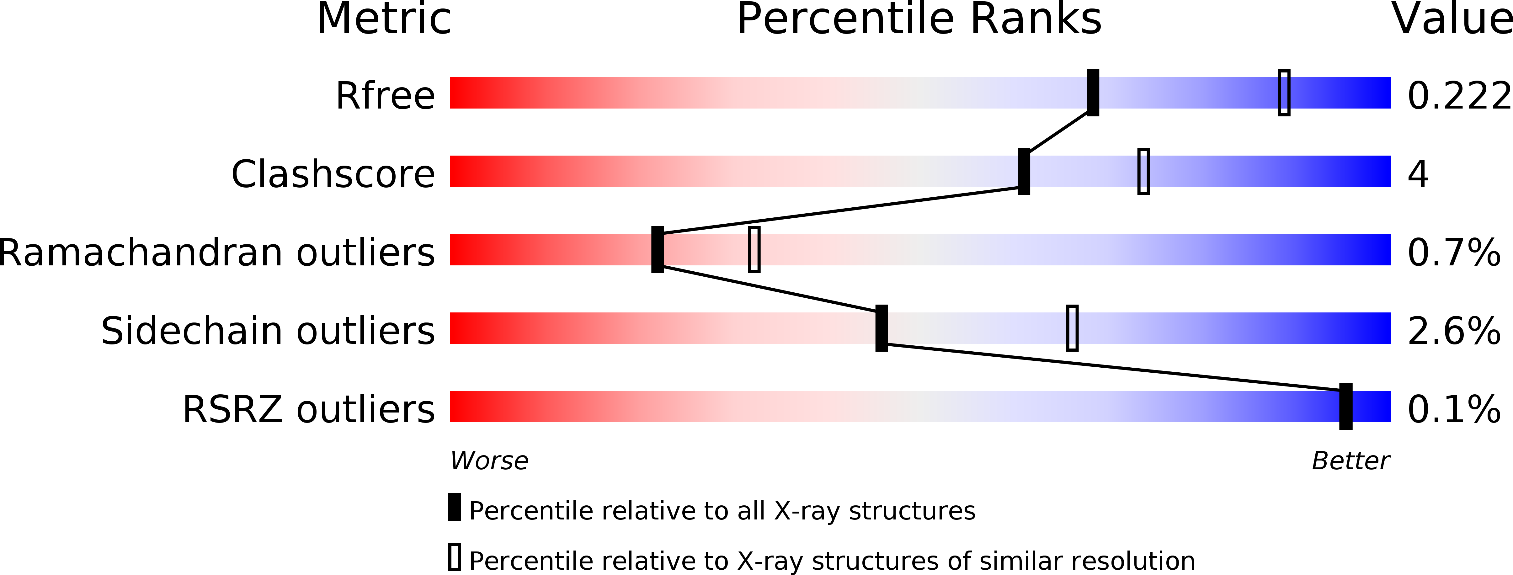
1ECE image
Entry Detail
PDB ID:
1ECE
Title:
ACIDOTHERMUS CELLULOLYTICUS ENDOCELLULASE E1 CATALYTIC DOMAIN IN COMPLEX WITH A CELLOTETRAOSE
Biological Source:
PDB Version:
Deposition Date:
1996-04-04
Release Date:
1996-10-14
Method Details:
Experimental Method:
Resolution:
2.40 Å
R-Value Free:
0.23
R-Value Work:
0.17
R-Value Observed:
0.17
Space Group:
P 32 2 1
Macromolecular Entities
Polymer Type:polypeptide(L)
Description:ENDOCELLULASE E1
Chain IDs:A, B
Chain Length:358
Number of Molecules:2
Biological Source:Acidothermus cellulolyticus
Peptide-like Molecules
PRD_900011
Primary Citation
Crystal structure of thermostable family 5 endocellulase E1 from Acidothermus cellulolyticus in complex with cellotetraose.
Biochemistry 35 10648 10660 (1996)
PMID: 8718854 DOI: 10.1021/bi9604439

Abstact

The crystal structure of the catalytic domain of the thermostable endocellulase E1 from Acidothermus cellulolyticus in complex with cellotetraose has been solved by multiple isomorphous replacement and refined at 2.4 A resolution to an R-factor of 0.18 (Rfree = 0.24). E1cd is a member of the 4/7 superfamily of hydrolases, and as expected, its structure is an (alpha/beta)8 barrel, which constitutes a prototype for family 5-subfamily 1 cellulases. The cellotetraose molecule binds in a manner consistent with the expected Michaelis complex for the glycosylation half-reaction and reveals that all eight residues conserved in family 5 enzymes are involved in recognition of the glycosyl group attacked during cleavage. Whereas only three residues are conserved in the whole 4/7 superfamily (the Asn/Glu duo and the Glu from which the name is derived), structural comparisons show that all eight residues conserved in family 5 have functional equivalents in the other 4/7 superfamily members, strengthening the case that mechanistic details are conserved throughout the superfamily. On the basis of the structure, a detailed sequence of physical steps of the cleavage mechanism is proposed. A close approach of two key glutamate residues provides an elegant mechanism for the shift in the pKa of the acid/base for the glycosylation and deglycosylation half-reactions. Finally, purely structural based comparisons are used to show that significant differences exist in structural similarity scores resulting from different methods and suggest that caution should be exercised in interpreting such results in terms of implied evolutional relationships.

Legend

Protein

Chemical

Disease

Primary Citation of related structures