1EC6 image
Deposition Date 2000-01-25
Release Date 2000-02-21
Last Version Date 2024-02-07
Entry Detail
PDB ID:
1EC6
Title:
CRYSTAL STRUCTURE OF NOVA-2 KH3 K-HOMOLOGY RNA-BINDING DOMAIN BOUND TO 20-MER RNA HAIRPIN
Biological Source:
Source Organism:
Homo sapiens (Taxon ID: 9606)
(Taxon ID: )
Host Organism:
Method Details:
Experimental Method:
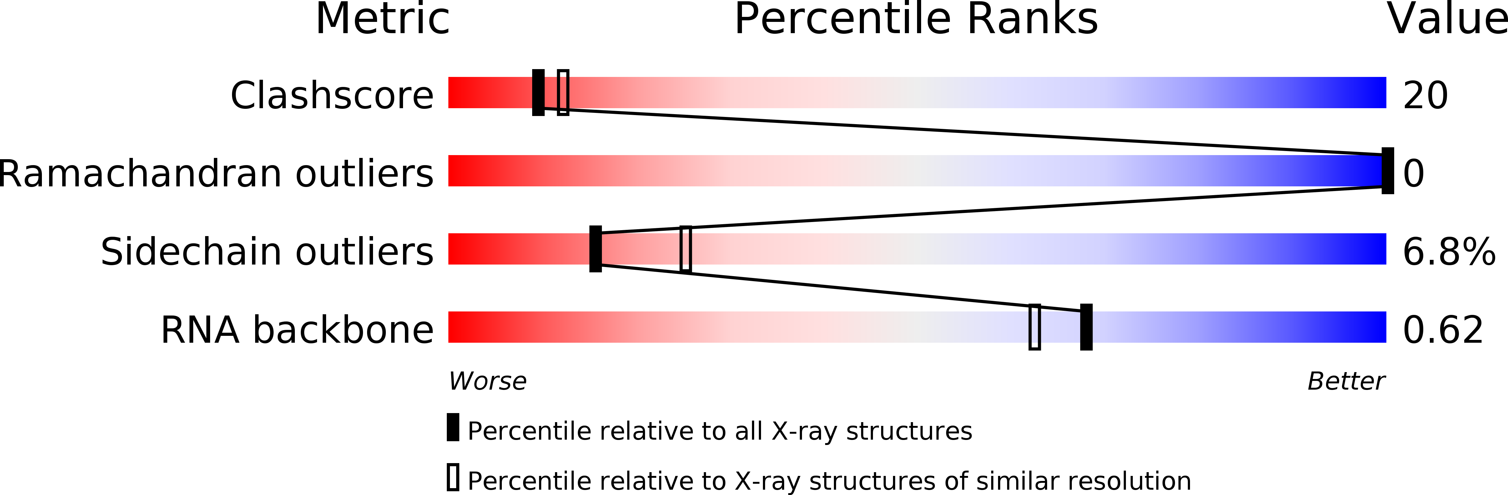
Resolution:
2.40 Å
R-Value Free:
0.29
R-Value Work:
0.20
R-Value Observed:
0.21
Space Group:
P 21 21 21
Macromolecular Entities
Polymer Type:polypeptide(L)
Molecule:RNA-BINDING PROTEIN NOVA-2
Gene (Uniprot):NOVA2
Chain IDs:C (auth: A), D (auth: B)
Chain Length:87
Number of Molecules:2
Biological Source:Homo sapiens
Polymer Type:polyribonucleotide
Molecule:20-MER RNA HAIRPIN
Chain IDs:A (auth: C), B (auth: D)
Chain Length:20
Number of Molecules:2
Biological Source:
Primary Citation
Sequence-specific RNA binding by a Nova KH domain: implications for paraneoplastic disease and the fragile X syndrome.
Cell(Cambridge,Mass.) 100 323 332 (2000)
PMID: 10676814 DOI: 10.1016/S0092-8674(00)80668-6

Abstact

The structure of a Nova protein K homology (KH) domain recognizing single-stranded RNA has been determined at 2.4 A resolution. Mammalian Nova antigens (1 and 2) constitute an important family of regulators of RNA metabolism in neurons, first identified using sera from cancer patients with the autoimmune disorder paraneoplastic opsoclonus-myoclonus ataxia (POMA). The structure of the third KH domain (KH3) of Nova-2 bound to a stem loop RNA resembles a molecular vise, with 5'-Ura-Cyt-Ade-Cyt-3' pinioned between an invariant Gly-X-X-Gly motif and the variable loop. Tetranucleotide recognition is supported by an aliphatic alpha helix/beta sheet RNA-binding platform, which mimics 5'-Ura-Gua-3' by making Watson-Crick-like hydrogen bonds with 5'-Cyt-Ade-3'. Sequence conservation suggests that fragile X mental retardation results from perturbation of RNA binding by the FMR1 protein.

Legend

Protein

Chemical

Disease

Primary Citation of related structures