
Deposition Date
1998-11-03
Release Date
1999-07-02
Last Version Date
2024-11-13
Entry Detail
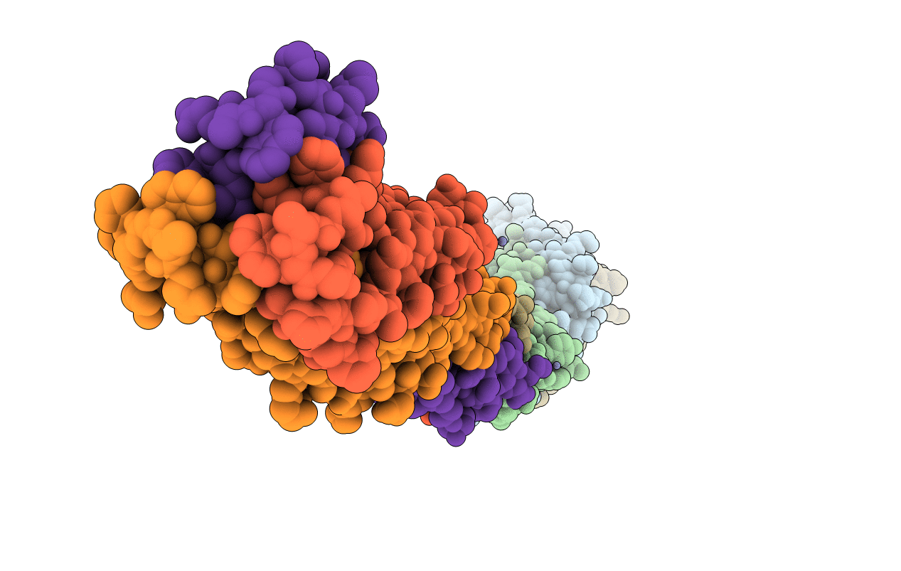
PDB ID:
1EBO
Keywords:
Title:
CRYSTAL STRUCTURE OF THE EBOLA VIRUS MEMBRANE-FUSION SUBUNIT, GP2, FROM THE ENVELOPE GLYCOPROTEIN ECTODOMAIN
Biological Source:
Source Organism(s):
Ebola virus sp. (Taxon ID: 205488)
Expression System(s):
Method Details:
Experimental Method:
Resolution:
3.00 Å
R-Value Free:
0.25
R-Value Work:
0.23
Space Group:
C 1 2 1
