1EBM image
Deposition Date 2000-01-24
Release Date 2000-03-20
Last Version Date 2024-02-07
Entry Detail
PDB ID:
1EBM
Keywords:
Title:
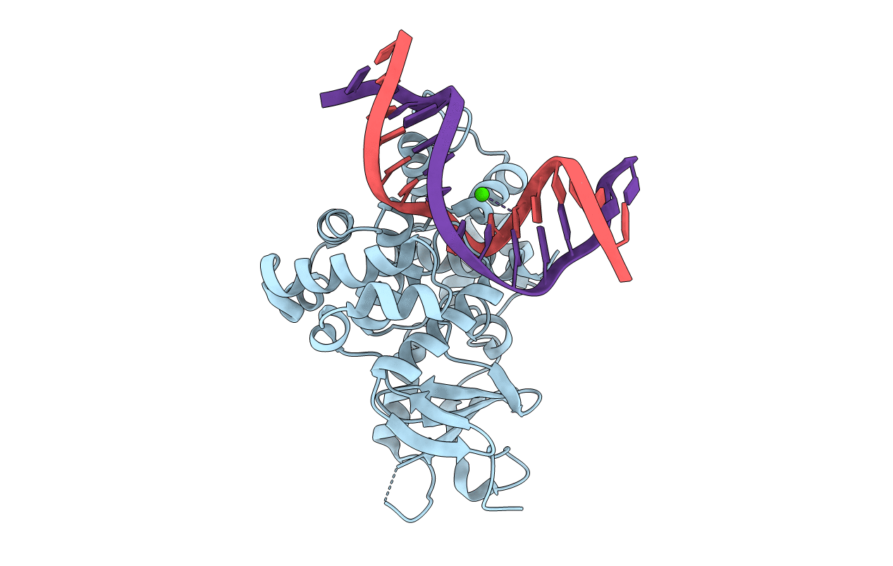
CRYSTAL STRUCTURE OF THE HUMAN 8-OXOGUANINE GLYCOSYLASE (HOGG1) BOUND TO A SUBSTRATE OLIGONUCLEOTIDE
Biological Source:
Source Organism:
Homo sapiens (Taxon ID: 9606)
Host Organism:
Method Details:
Experimental Method:
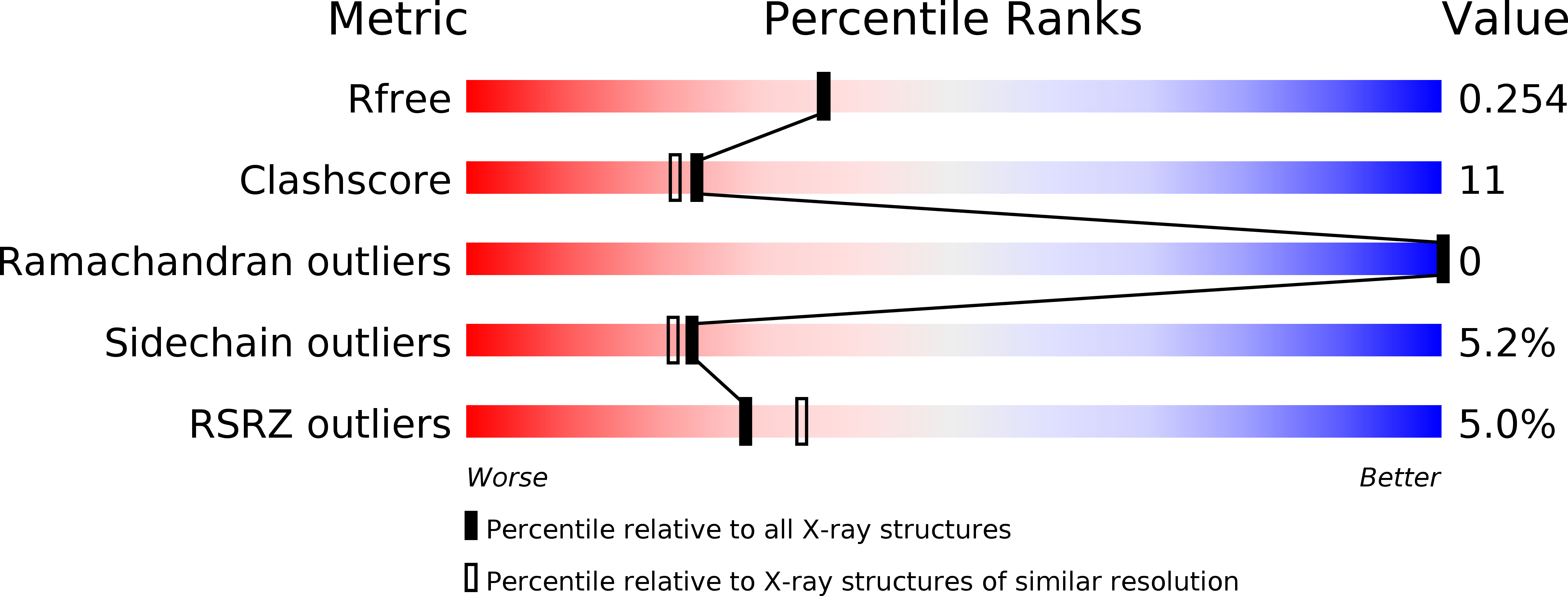
Resolution:
2.10 Å
R-Value Free:
0.26
R-Value Work:
0.23
R-Value Observed:
0.23
Space Group:
P 65 2 2
Macromolecular Entities
Polymer Type:polypeptide(L)
Molecule:8-OXOGUANINE DNA GLYCOSYLASE
Gene (Uniprot):OGG1
Mutations:K249Q
Chain IDs:C (auth: A)
Chain Length:317
Number of Molecules:1
Biological Source:Homo sapiens
Modified Residue
Compound ID Chain ID Parent Comp ID Details 2D Image
8OG A DG ?
Ligand Molecules
Primary Citation
Structural basis for recognition and repair of the endogenous mutagen 8-oxoguanine in DNA.
Nature 403 859 866 (2000)
PMID: 10706276 DOI: 10.1038/35002510

Abstact

Spontaneous oxidation of guanine residues in DNA generates 8-oxoguanine (oxoG). By mispairing with adenine during replication, oxoG gives rise to a G x C --> T x A transversion, a frequent somatic mutation in human cancers. The dedicated repair pathway for oxoG centres on 8-oxoguanine DNA glycosylase (hOGG1), an enzyme that recognizes oxoG x C base pairs, catalysing expulsion of the oxoG and cleavage of the DNA backbone. Here we report the X-ray structure of the catalytic core of hOGG1 bound to oxoG x C-containing DNA at 2.1 A resolution. The structure reveals the mechanistic basis for the recognition and catalytic excision of DNA damage by hOGG1 and by other members of the enzyme superfamily to which it belongs. The structure also provides a rationale for the biochemical effects of inactivating mutations and polymorphisms in hOGG1. One known mutation, R154H, converts hOGG1 to a promutator by relaxing the specificity of the enzyme for the base opposite oxoG.

Legend

Protein

Chemical

Disease

Primary Citation of related structures