1EBF image
Deposition Date 2000-01-24
Release Date 2000-03-08
Last Version Date 2024-02-07
Entry Detail
PDB ID:
1EBF
Keywords:
Title:
HOMOSERINE DEHYDROGENASE FROM S. CEREVISIAE COMPLEX WITH NAD+
Biological Source:
Source Organism:
Host Organism:
Method Details:
Experimental Method:
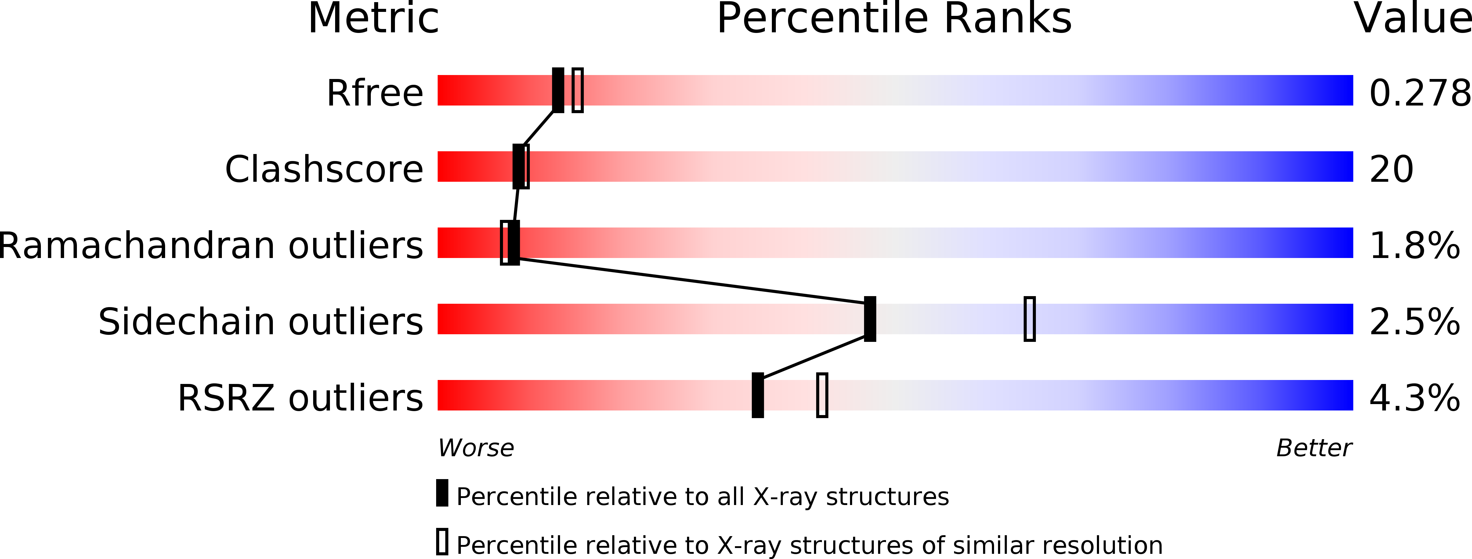
Resolution:
2.30 Å
R-Value Free:
0.26
R-Value Work:
0.21
R-Value Observed:
0.21
Space Group:
P 43 21 2
Macromolecular Entities
Structures with similar UniProt ID
Protein Blast
Polymer Type:polypeptide(L)
Molecule:HOMOSERINE DEHYDROGENASE
Gene (Uniprot):HOM6
Chain IDs:A, B
Chain Length:358
Number of Molecules:2
Biological Source:Saccharomyces cerevisiae
Primary Citation
Crystal structures of homoserine dehydrogenase suggest a novel catalytic mechanism for oxidoreductases.
Nat.Struct.Biol. 7 238 244 (2000)
PMID: 10700284 DOI: 10.1038/73359

Abstact

The structure of the antifungal drug target homoserine dehydrogenase (HSD) was determined from Saccharomyces cerevisiae in apo and holo forms, and as a ternary complex with bound products, by X-ray diffraction. The three forms show that the enzyme is a dimer, with each monomer composed of three regions, the nucleotide-binding region, the dimerization region and the catalytic region. The dimerization and catalytic regions have novel folds, whereas the fold of the nucleotide-binding region is a variation on the Rossmann fold. The novel folds impose a novel composition and arrangement of active site residues when compared to all other currently known oxidoreductases. This observation, in conjunction with site-directed mutagenesis of active site residues and steady-state kinetic measurements, suggest that HSD exhibits a new variation on dehydrogenase chemistry.

Legend

Protein

Chemical

Disease

Primary Citation of related structures