Deposition Date
1996-02-03
Release Date
1996-07-11
Last Version Date
2024-10-23
Entry Detail
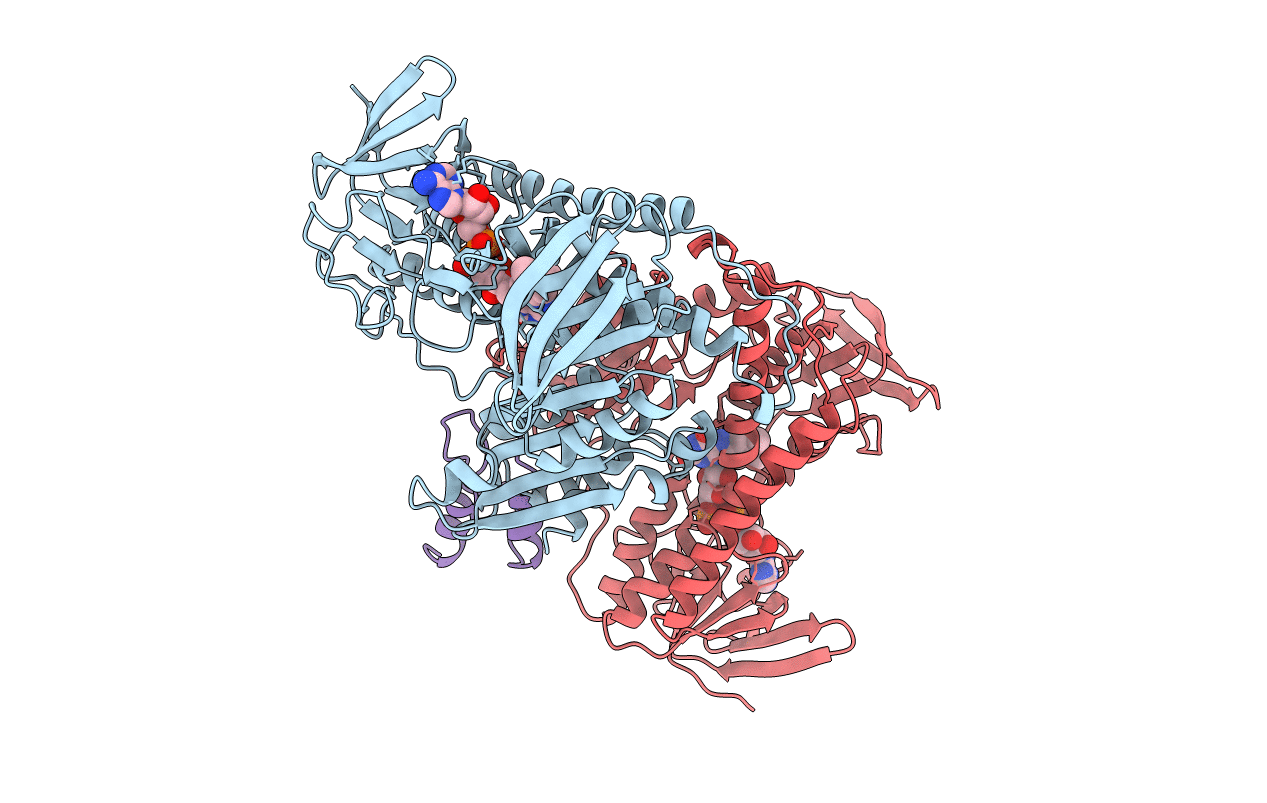
PDB ID:
1EBD
Keywords:
Title:
DIHYDROLIPOAMIDE DEHYDROGENASE COMPLEXED WITH THE BINDING DOMAIN OF THE DIHYDROLIPOAMIDE ACETYLASE
Biological Source:
Source Organism(s):
Geobacillus stearothermophilus (Taxon ID: 1422)
Expression System(s):
Method Details:
Experimental Method:
Resolution:
2.60 Å
R-Value Work:
0.21
R-Value Observed:
0.21
Space Group:
P 31 2 1