1EAV image
Deposition Date 2001-07-17
Release Date 2001-11-23
Last Version Date 2024-11-20
Entry Detail
PDB ID:
1EAV
Title:
Crystal Structures of Human Gephyrin and Plant Cnx1 G domains - Comparative Analysis and Functional Implications
Biological Source:
Source Organism:
Host Organism:
Method Details:
Experimental Method:
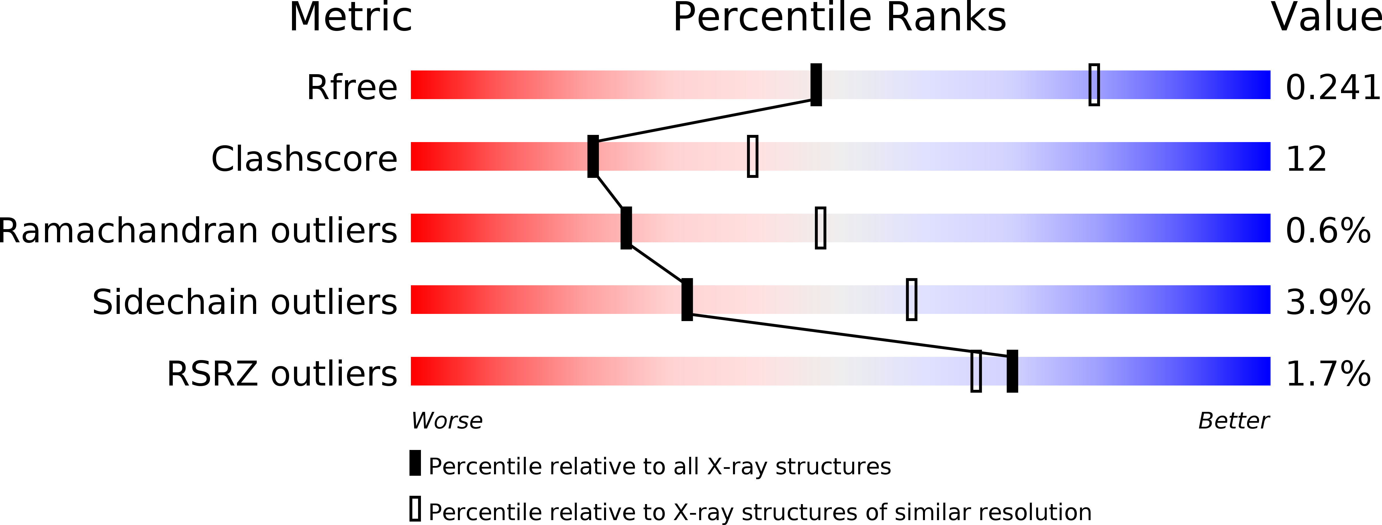
Resolution:
2.60 Å
R-Value Free:
0.25
R-Value Work:
0.22
Space Group:
P 21 3
Macromolecular Entities
Polymer Type:polypeptide(L)
Molecule:MOLYBDOPTERIN BIOSYNTHESIS CNX1 PROTEIN
Gene (Uniprot):CNX1
Chain IDs:A, B, C, D, E, F, G, H
Chain Length:162
Number of Molecules:8
Biological Source:ARABIDOPSIS THALIANA
Modified Residue
Compound ID Chain ID Parent Comp ID Details 2D Image
MSE A MET SELENOMETHIONINE
Primary Citation
Crystal Structures of Human Gephyrin and Plant Cnx1 G Domains: Comparative Analysis and Functional Implications
J.Mol.Biol. 312 405 ? (2001)
PMID: 11554796 DOI: 10.1006/JMBI.2001.4952

Abstact

The molybdenum cofactor (Moco) consists of a unique and conserved pterin derivative, usually referred to as molybdopterin (MPT), which coordinates the essential transition metal molybdenum (Mo). Moco is required for the enzymatic activities of all Mo-enzymes, with the exception of nitrogenase and is synthesized by an evolutionary old multi-step pathway that is dependent on the activities of at least six gene products. In eukaryotes, the final step of Moco biosynthesis, i.e. transfer and insertion of Mo into MPT, is catalyzed by the two-domain proteins Cnx1 in plants and gephyrin in mammals. Gephyrin is ubiquitously expressed, and was initially found in the central nervous system, where it is essential for clustering of inhibitory neuroreceptors in the postsynaptic membrane. Gephyrin and Cnx1 contain at least two functional domains (E and G) that are homologous to the Escherichia coli proteins MoeA and MogA, the atomic structures of which have been solved recently. Here, we present the crystal structures of the N-terminal human gephyrin G domain (Geph-G) and the C-terminal Arabidopsis thaliana Cnx1 G domain (Cnx1-G) at 1.7 and 2.6 A resolution, respectively. These structures are highly similar and compared to MogA reveal four major differences in their three-dimensional structures: (1) In Geph-G and Cnx1-G an additional alpha-helix is present between the first beta-strand and alpha-helix of MogA. (2) The loop between alpha 2 and beta 2 undergoes conformational changes in all three structures. (3) A beta-hairpin loop found in MogA is absent from Geph-G and Cnx1-G. (4) The C terminus of Geph-G follows a different path from that in MogA. Based on the structures of the eukaryotic proteins and their comparisons with E. coli MogA, the predicted binding site for MPT has been further refined. In addition, the characterized alternative splice variants of gephyrin are analyzed in the context of the three-dimensional structure of Geph-G.

Legend

Protein

Chemical

Disease

Primary Citation of related structures