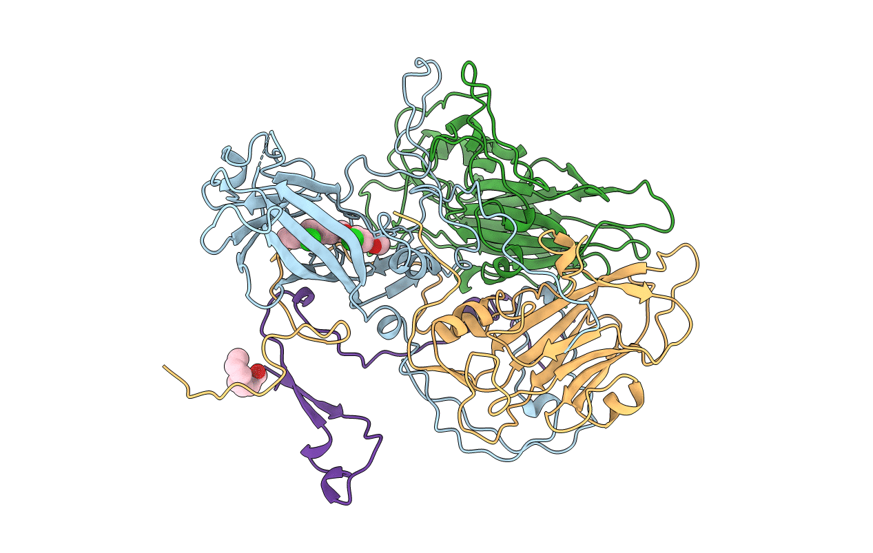
1EAH image
Entry Detail
PDB ID:
1EAH
Keywords:
Title:
PV2L COMPLEXED WITH ANTIVIRAL AGENT SCH48973
Biological Source:
Source Organism:
Host Organism:
PDB Version:
Deposition Date:
1997-07-22
Release Date:
1998-09-16
Method Details:
Experimental Method:
Resolution:
2.90 Å
R-Value Work:
0.18
R-Value Observed:
0.18
Space Group:
C 2 2 21
Macromolecular Entities
Polymer Type:polypeptide(L)
Description:POLIOVIRUS TYPE 2 COAT PROTEINS VP1 TO VP4
Chain IDs:A (auth: 1)
Chain Length:301
Number of Molecules:1
Biological Source:Human poliovirus 2
Polymer Type:polypeptide(L)
Description:POLIOVIRUS TYPE 2 COAT PROTEINS VP1 TO VP4
Chain IDs:B (auth: 2)
Chain Length:271
Number of Molecules:1
Biological Source:Human poliovirus 2
Polymer Type:polypeptide(L)
Description:POLIOVIRUS TYPE 2 COAT PROTEINS VP1 TO VP4
Chain IDs:C (auth: 3)
Chain Length:238
Number of Molecules:1
Biological Source:Human poliovirus 2
Polymer Type:polypeptide(L)
Description:POLIOVIRUS TYPE 2 COAT PROTEINS VP1 TO VP4
Chain IDs:D (auth: 4)
Chain Length:68
Number of Molecules:1
Biological Source:Human poliovirus 2
Primary Citation

Abstact

BACKGROUND Polioviruses are human pathogens and the causative agents of poliomyelitis. Polioviruses are icosahedral single-stranded RNA viruses, which belong to the picornavirus family, and occur as three distinct serotypes. All three serotypes of poliovirus can infect primates, but only type 2 can infect mice. The crystal structures of a type 1 and a type 3 poliovirus are already known. Structural studies of poliovirus type 2 Lansing (PV2L) were initiated to try to enhance our understanding of the differences in host range specificity, antigenicity and receptor binding among the three serotypes of poliovirus. RESULTS The crystal structure of the mouse neurovirulent PV2L complexed with a potent antiviral agent, SCH48973, was determined at 2.9 A resolution. Structural differences among the three poliovirus serotypes occur primarily in the loop regions of the viral coat proteins (VPs), most notably in the loops of VP1 that cluster near the fivefold axes of the capsid, where the BC loop of PV2L is disordered. Unlike other known structures of enteroviruses, the entire polypeptide chain of PV2L VP4 is visible in the electron density and RNA bases are observed stacking with conserved aromatic residues (Tyr4020 and Phe4046) of VP4. The broad-spectrum antiviral agent SCH48973 is observed binding in a pocket within the beta-barrel of VP1, in approximately the same location that natural 'pocket factors' bind to polioviruses. SCH48973 forms predominantly hydrophobic interactions with the pocket residues. CONCLUSIONS Some of the conformational changes required for infectivity and involved in the control of capsid stability and neurovirulence in mice may occur in the vicinity of the fivefold axis of the poliovirus, where there are significant structural differences among the three poliovirus serotypes in the surface exposed loops of VP1 (BC, DE, and HI). A surface depression is located at the fivefold axis of PV2L that is not present in the other two poliovirus serotypes. The observed interaction of RNA with VP4 supports the observation that loss of VP4 ultimately leads to the loss of viral RNA. A model is proposed that suggests dual involvement of the virion fivefold and pseudo-threefold axes in receptor-mediated initiation of infection by picornaviruses.

Legend

Protein

Chemical

Disease

Primary Citation of related structures