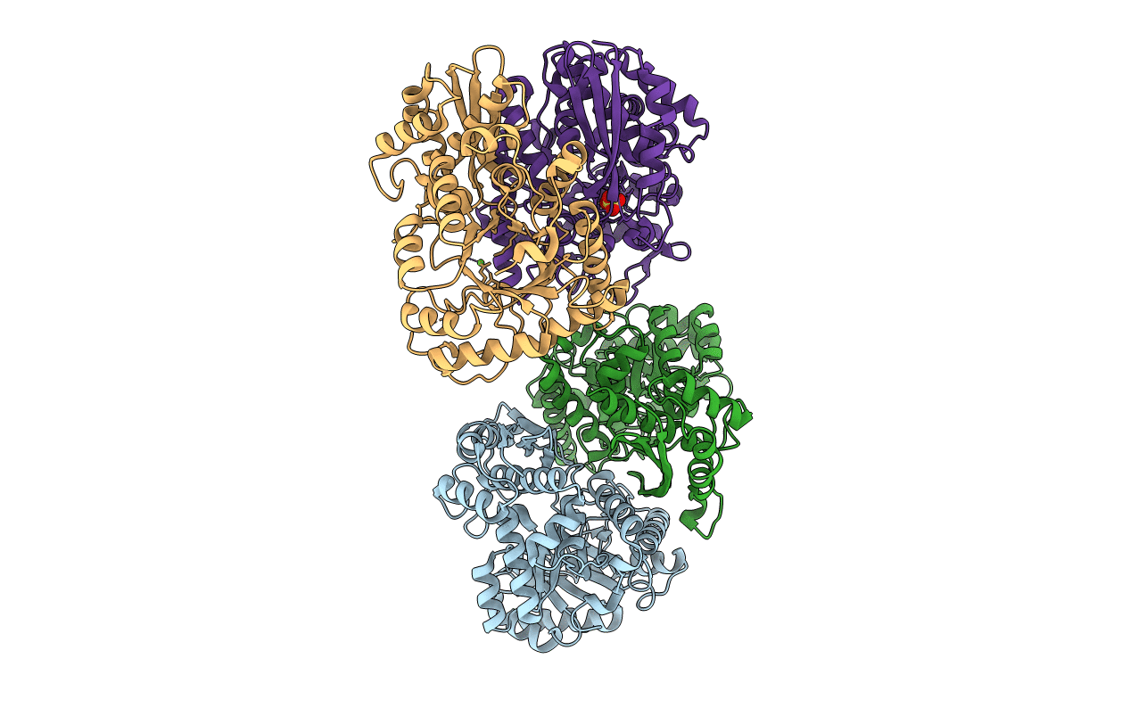
1E9I image
Deposition Date 2000-10-17
Release Date 2001-03-15
Last Version Date 2023-12-13
Entry Detail
PDB ID:
1E9I
Keywords:
Title:
Enolase from E.coli
Biological Source:
Source Organism:
ESCHERICHIA COLI (Taxon ID: 562)
Host Organism:
Method Details:
Experimental Method:
Resolution:
2.48 Å
R-Value Free:
0.27
R-Value Work:
0.22
R-Value Observed:
0.22
Space Group:
C 1 2 1
Macromolecular Entities
Polymer Type:polypeptide(L)
Molecule:ENOLASE
Chain IDs:A, B, C, D
Chain Length:431
Number of Molecules:4
Biological Source:ESCHERICHIA COLI
Primary Citation
Crystal Structure of the Escherichia Coli RNA Degradosome Component Enolase
J.Mol.Biol. 313 583 ? (2001)
PMID: 11676541 DOI: 10.1006/JMBI.2001.5065

Abstact

The crystal structure of Escherichia coli enolase (EC 4.2.1.11, phosphopyruvate hydratase), which is a component of the RNA degradosome, has been determined at 2.5 A. There are four molecules in the asymmetric unit of the C2 cell, and in one of the molecules, flexible loops close onto the active site. This closure mimics the conformation of the substrate-bound intermediate. A comparison of the structure of the E. coli enolase with the eukaryotic enolase structures available (lobster and yeast) indicates a high degree of conservation of the hydrophobic core and the subunit interface of this homodimeric enzyme. The dimer interface is enriched in charged residues compared with other protein homodimers, which may explain our observations from analytical ultracentrifugation that dimerisation is affected by ionic strength. The putative role of enolase in the RNA degradosome is discussed; although it was not possible to ascribe a specific role to it, a structural role is possible.

Legend

Protein

Chemical

Disease

Primary Citation of related structures