1E8C image
Entry Detail
PDB ID:
1E8C
Keywords:
Title:
Structure of MurE the UDP-N-acetylmuramyl tripeptide synthetase from E. coli
Biological Source:
Source Organism:
Host Organism:
PDB Version:
Deposition Date:
2000-09-19
Release Date:
2001-09-13
Method Details:
Experimental Method:
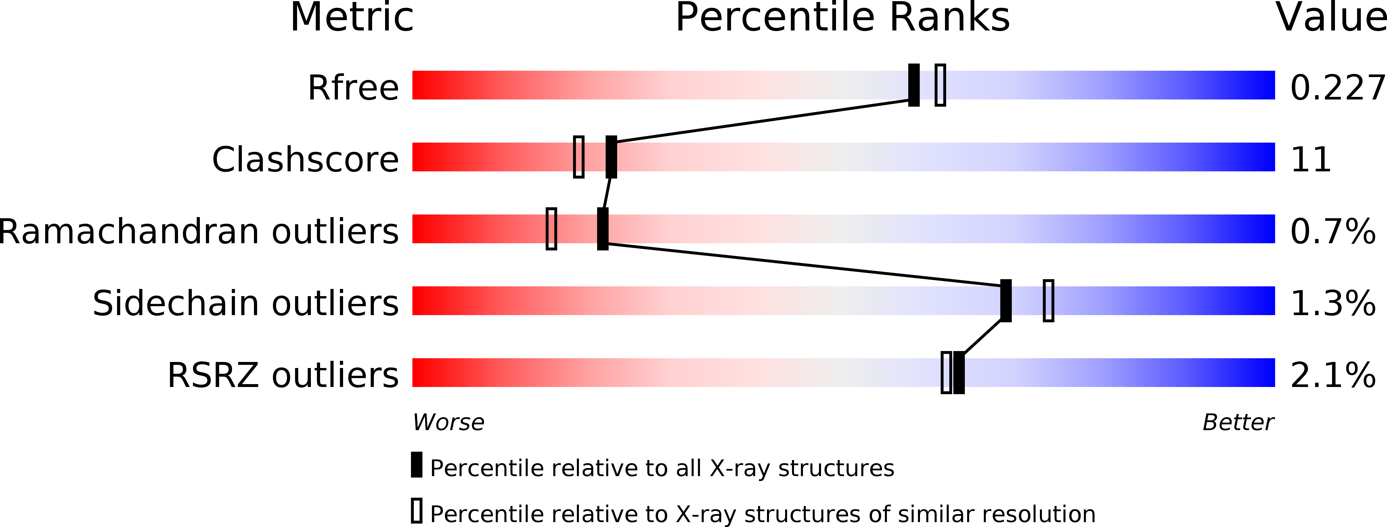
Resolution:
2.00 Å
R-Value Free:
0.23
R-Value Work:
0.20
R-Value Observed:
0.20
Space Group:
C 2 2 21
Macromolecular Entities
Polymer Type:polypeptide(L)
Description:UDP-N-ACETYLMURAMOYLALANYL-D-GLUTAMATE--2,6-DIAMINOPIMELATE LIGASE
Chain IDs:A, B
Chain Length:498
Number of Molecules:2
Biological Source:ESCHERICHIA COLI
Modified Residue
Compound ID Chain ID Parent Comp ID Details 2D Image
KCX A LYS LYSINE NZ-CARBOXYLIC ACID
MSE A MET SELENOMETHIONINE
Primary Citation
Crystal Structure of Udp-N-Acetylmuramoyl-L-Alanyl-D-Glutamate: Meso-Diaminopimelate Ligase from Escherichia Coli
J.Biol.Chem. 276 10999 ? (2001)
PMID: 11124264 DOI: 10.1074/JBC.M009835200

Abstact

UDP-N-acetylmuramoyl-l-alanyl-d-glutamate:meso-diaminopimelate ligase is a cytoplasmic enzyme that catalyzes the addition of meso-diaminopimelic acid to nucleotide precursor UDP-N-acetylmuramoyl-l-alanyl-d-glutamate in the biosynthesis of bacterial cell-wall peptidoglycan. The crystal structure of the Escherichia coli enzyme in the presence of the final product of the enzymatic reaction, UDP-MurNAc-l-Ala-gamma-d-Glu-meso-A(2)pm, has been solved to 2.0 A resolution. Phase information was obtained by multiwavelength anomalous dispersion using the K shell edge of selenium. The protein consists of three domains, two of which have a topology reminiscent of the equivalent domain found in the already established three-dimensional structure of the UDP-N-acetylmuramoyl-l-alanine: D-glutamate-ligase (MurD) ligase, which catalyzes the immediate previous step of incorporation of d-glutamic acid in the biosynthesis of the peptidoglycan precursor. The refined model reveals the binding site for UDP-MurNAc-l-Ala-gamma-d-Glu-meso-A(2)pm, and comparison with the six known MurD structures allowed the identification of residues involved in the enzymatic mechanism. Interestingly, during refinement, an excess of electron density was observed, leading to the conclusion that, as in MurD, a carbamylated lysine residue is present in the active site. In addition, the structural determinant responsible for the selection of the amino acid to be added to the nucleotide precursor was identified.

Legend

Protein

Chemical

Disease

Primary Citation of related structures