1E6Y image
Deposition Date 2000-08-23
Release Date 2000-10-18
Last Version Date 2023-12-13
Entry Detail
PDB ID:
1E6Y
Keywords:
Title:
Methyl-coenzyme M reductase from Methanosarcina barkeri
Biological Source:
Source Organism:
Method Details:
Experimental Method:
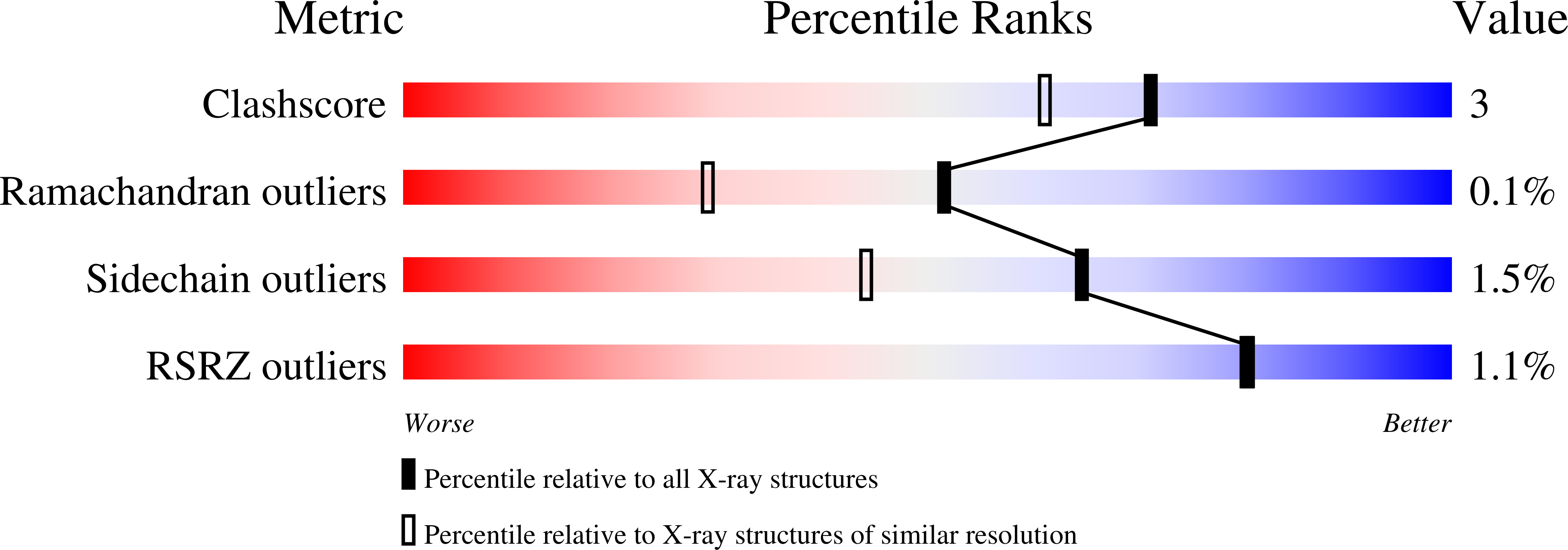
Resolution:
1.60 Å
R-Value Free:
0.17
R-Value Work:
0.16
R-Value Observed:
0.16
Space Group:
P 21 21 21
Macromolecular Entities
Structures with similar UniProt ID
Protein Blast
Polymer Type:polypeptide(L)
Molecule:METHYL-COENZYME M REDUCTASE SUBUNIT ALPHA
Gene (Uniprot):mcrA
Chain IDs:A, D
Chain Length:569
Number of Molecules:2
Biological Source:METHANOSARCINA BARKERI
Structures with similar UniProt ID
Protein Blast
Polymer Type:polypeptide(L)
Molecule:METHYL-COENZYME M REDUCTASE I BETA SUBUNIT
Gene (Uniprot):mcrB
Chain IDs:B, E
Chain Length:433
Number of Molecules:2
Biological Source:METHANOSARCINA BARKERI
Structures with similar UniProt ID
Protein Blast
Polymer Type:polypeptide(L)
Molecule:METHYL-COENZYME M REDUCTASE SUBUNIT GAMMA
Gene (Uniprot):mcrG
Chain IDs:C, F
Chain Length:247
Number of Molecules:2
Biological Source:METHANOSARCINA BARKERI
Modified Residue
Compound ID Chain ID Parent Comp ID Details 2D Image
AGM A ARG 5-METHYL-ARGININE
GL3 A GLY THIOGLYCIN
MHS A HIS N1-METHYLATED HISTIDINE
OCS C CYS CYSTEINESULFONIC ACID
SMC A CYS S-METHYLCYSTEINE
Primary Citation
Comparison of Three Methyl-Coenzyme M Reductases from Phylogenetically Distant Organisms: Unusual Amino Acid Modification, Conservation and Adaptation
J.Mol.Biol. 303 329 ? (2000)
PMID: 11023796 DOI: 10.1006/JMBI.2000.4136

Abstact

The nickel enzyme methyl-coenzyme M reductase (MCR) catalyzes the terminal step of methane formation in the energy metabolism of all methanogenic archaea. In this reaction methyl-coenzyme M and coenzyme B are converted to methane and the heterodisulfide of coenzyme M and coenzyme B. The crystal structures of methyl-coenzyme M reductase from Methanosarcina barkeri (growth temperature optimum, 37 degrees C) and Methanopyrus kandleri (growth temperature optimum, 98 degrees C) were determined and compared with the known structure of MCR from Methanobacterium thermoautotrophicum (growth temperature optimum, 65 degrees C). The active sites of MCR from M. barkeri and M. kandleri were almost identical to that of M. thermoautotrophicum and predominantly occupied by coenzyme M and coenzyme B. The electron density at 1.6 A resolution of the M. barkeri enzyme revealed that four of the five modified amino acid residues of MCR from M. thermoautotrophicum, namely a thiopeptide, an S-methylcysteine, a 1-N-methylhistidine and a 5-methylarginine were also present. Analysis of the environment of the unusual amino acid residues near the active site indicates that some of the modifications may be required for the enzyme to be catalytically effective. In M. thermoautotrophicum and M. kandleri high temperature adaptation is coupled with increasing intracellular concentrations of lyotropic salts. This was reflected in a higher fraction of glutamate residues at the protein surface of the thermophilic enzymes adapted to high intracellular salt concentrations.

Legend

Protein

Chemical

Disease

Primary Citation of related structures
Feedback Form
Name
Email
Institute
Feedback