1E6J image
Deposition Date 2000-08-18
Release Date 2000-11-27
Last Version Date 2024-10-16
Entry Detail
PDB ID:
1E6J
Keywords:
Title:
Crystal structure of HIV-1 capsid protein (p24) in complex with Fab13B5
Biological Source:
Source Organism:
HIV-1 M\:B_HXB2R (Taxon ID: 11706)
MUS MUSCULUS (Taxon ID: 10090)
Host Organism:
Method Details:
Experimental Method:
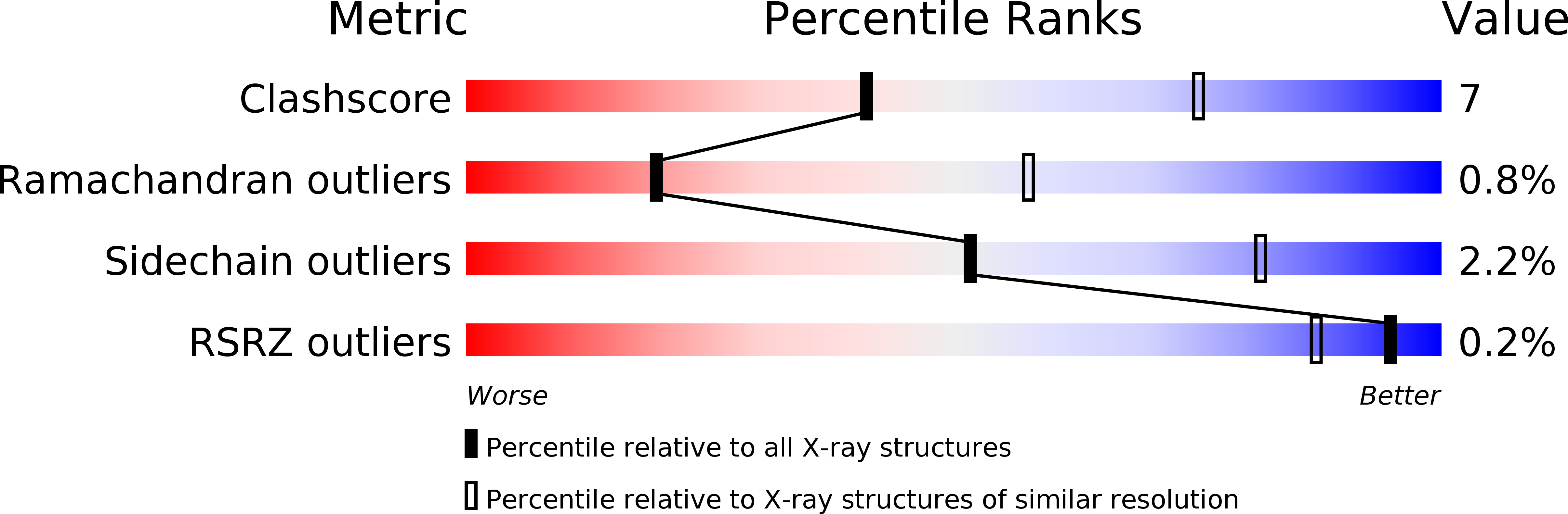
Resolution:
3.00 Å
R-Value Free:
0.28
R-Value Work:
0.21
R-Value Observed:
0.21
Space Group:
C 1 2 1
Macromolecular Entities
Polymer Type:polypeptide(L)
Molecule:IMMUNOGLOBULIN
Chain IDs:A (auth: H)
Chain Length:219
Number of Molecules:1
Biological Source:MUS MUSCULUS
Polymer Type:polypeptide(L)
Molecule:IMMUNOGLOBULIN
Chain IDs:B (auth: L)
Chain Length:210
Number of Molecules:1
Biological Source:MUS MUSCULUS
Polymer Type:polypeptide(L)
Molecule:CAPSID PROTEIN P24
Chain IDs:C (auth: P)
Chain Length:210
Number of Molecules:1
Biological Source:HIV-1 M\:B_HXB2R
Ligand Molecules
Primary Citation
Mutual Conformational Adaptations in Antigen and Antibody Upon Complex Formation between an Fab and HIV-1 Capsid Protein P24
Structure 8 1069 ? (2000)
PMID: 11080628 DOI: 10.1016/S0969-2126(00)00507-4

Abstact

BACKGROUND: Elucidating the structural basis of antigen-antibody recognition ideally requires a structural comparison of free and complexed components. To this end we have studied a mouse monoclonal antibody, denoted 13B5, raised against p24, the capsid protein of HIV-1. We have previously described the first crystal structure of intact p24 as visualized in the Fab13B5-p24 complex. Here we report the structure of the uncomplexed Fab13B5 at 1.8 A resolution and analyze the Fab-p24 interface and the conformational changes occurring upon complex formation. RESULTS: Fab13B5 recognizes a nearly continuous epitope comprising a helix-turn-helix motif in the C-terminal domain of p24. Only 4 complementarity-determining regions (CDRs) are in contact with p24 with most interactions being by the heavy chain. Comparison of the free and complexed Fab reveals that structural changes upon binding are localized to a few side chains of CDR-H1 and -H2 but involve a larger, concerted displacement of CDR-H3. Antigen binding is also associated with an 8 degrees relative rotation of the heavy and light chain variable regions. In p24, small conformational changes localized to the turn between the two helices comprising the epitope result from Fab binding. CONCLUSIONS: The relatively small area of contact between Fab13B5 and p24 may be related to the fact that the epitope is a continuous peptide rather than a more complex protein surface and correlates with a relatively low affinity of antigen and antibody. Despite this, a significant quaternary structural change occurs in the Fab upon complex formation, with additional smaller adaptations of both antigen and antibody.

Legend

Protein

Chemical

Disease

Primary Citation of related structures